Vítor Cavadas graduated from medical school at the Faculty of Medicine of Oporto in 2002. He did his general internship at the São João Hospital in Oporto between 2003 and 2004 and completed his residency in Urology at the Santo António General Hospital in Oporto between 2005 and 2010. He became a consultant at the Department of Urology of the Centro Hospitalar Universitário do Porto in 2011 and has since been responsible for the Stone Unit in this Department. He is also member of Transplantion team in the same institution. He is particularly interested in stone disease management and upper tract tumours conservative treatment. He is a member of the Portuguese Association of Urology (leading the Lithiasis Working Group), the European Association of Urology and the Societé Internationale d’Urologie. He has participated in clinical trials and authored several articles in peer-reviewed journals. He frequently organizes courses on stone treatment and participates as surgeon in Live Surgery events. He has been awarded a research grant by the Portuguese Association of Urology and is also involved in the pre-graduate teaching of Urology at the Institute of Biomedical Sciences Abel Salazar in Oporto.
Selected publications from PubMed
Challenges in Follow-up After Kidney-sparing Surgery for Upper Tract Urothelial Carcinoma: Insights from a Delphi Consensus.
Figaroa O, Schuil H, Kamphuis G, van den Brink L, Hendriks N, Henderickx M, Bins A, van Moorselaar J, Ajayi L, Bus M, Cavadas V, Cracco CM, Andrea DD, Dasgupta R, Coninck V, Durutovic O, Ebbensgaard I, Ferretti S, Gallioli A, Keller EX, Keeley FX Jr, Sloth Osther PJ, Popiolek M, Pradere B, Proietti S, Saltirov I, Somani B, Tailly T, Thomas K, Turney B, Ulvik Ø, Haute CV, van der Heij B, Vasquez JL, Brehmer M, Baard J
Eur Urol Oncol. 2025 Oct;8(5):1303-1310 doi: 10.1016/j.euo.2025.07.003.
Stone-event-free survival after retrograde intrarenal surgery: is the stone-free-status so relevant for the future outcomes?
Mesquita S, Mendes G, Marques-Monteiro M, Rocha MA, Madanelo M, Fraga A, Cavadas V
Int Urol Nephrol. 2025 May;57(5):1473-1480 doi: 10.1007/s11255-024-04343-8.
Kidney stone analysis: an EAU section of urolithiasis (EULIS) survey on current practices and perspectives worldwide.
Stoots SJM, Somani BK, Durutovic O, Cavadas V, Secker A, Jung HU, Ulvik Ø, Ingimarsson J, Tefik T, Sener E, Dragos L, Tailly T, Popiolek M, Hamri SB, Gauhar V, M Zeeshan Hameed B, Rivas JG, Bhojani N, Seitz C, Beerlage H, de Jonge R, Kamphuis GM
World J Urol. 2024 Dec 10;43(1):21 doi: 10.1007/s00345-024-05348-9.
Should patients with encrusted JJ stents involving the proximal/renal loop undergo primarily endoscopic combined intrarenal surgery?
Rocha A, Mendes G, Mesquita S, Madanelo M, Vital J, Marques-Monteiro M, Vinagre N, Magalhães M, Oliveira B, Gonçalves G, Cavadas V, Fraga A
Arch Ital Urol Androl. 2024 Nov 11;96(4):13163 doi: 10.4081/aiua.2024.13163.
Nephrectomy with Autotransplantation-A Key Treasure.
Mesquita S, Marques-Monteiro M, Madanelo M, Rocha MA, Vinagre N, Fraga A, Cavadas V, Machado R, Silva-Ramos M
J Clin Med. 2024 Mar 13;13(6) doi: 10.3390/jcm13061641.
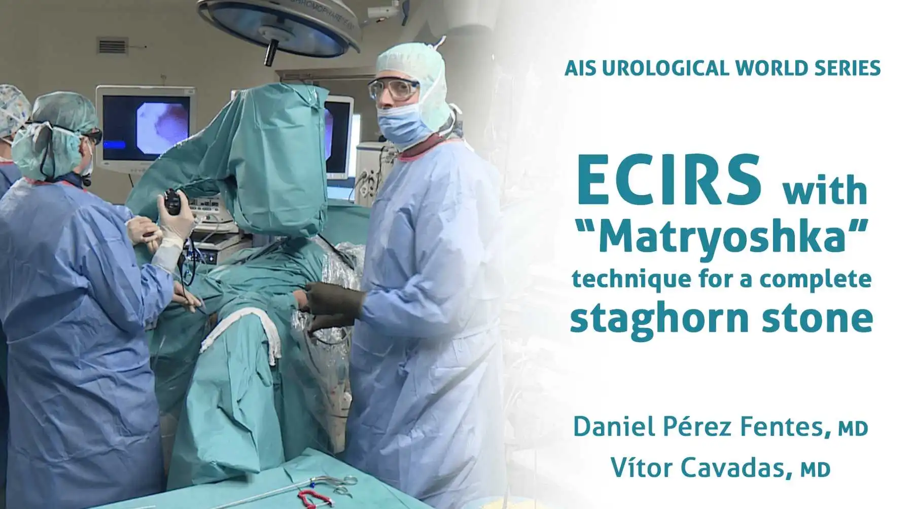
The patient was placed in the Galdakao-modified supine Valdivia position. A puncture was made under ultrasound and X-ray control in a posterior lower calyx, seeking a transpapillary puncture. The calyx entry was blocked, and the guidewire could not pass into the renal pelvis. An initial dilation of up to 16Ch was required to make room in this calyx access so to then place the guidewire in the ureter (“through and through” situation). A standard access (24Ch) was created using Amplatz serial dilators. A rigid nephroscope and lithotripsy using combined ultrasonic and ballistic energy with suction was used to remove the stone burden in the calyx access and renal pelvis. Flexible nephroscopy and laser were required to treat some fragments in the upper calyx and proximal ureter. Stones in the middle calyx were cleared out using a flexible ureteroscope inserted retrogradely (ECIRS) and extracted through the Amplatz sheath. A final endoscopic review did not reveal any residual fragments. At the end of the procedure a double J stent was placed without a nephrostomy tube, after having performed an endoscopic control of the percutaneous tract.
With Boston Scientific
Published
Jul 2019

Patient was placed in the Galdakao-modified supine Valdivia position. The double J stent was removed and a semirigid ureteroscopy was carried out immediately after. The ureteral stone was treated with Holmium laser and the fragments were actively removed using nitinol baskets. After having cleared out the ureter, a ureteral access sheath was inserted and a single-use flexible ureteroscope (LithoVueTM) was used to inspect the pelvicalyceal system. A percutaneous renal puncture was carried out under ultrasound and endovision control in a posterior lower calyx, looking for a transpapillary puncture. The tract was dilated up to 16Ch under endoscopic control. The different stones were treated using Holmium laser either through the rigid nephroscope and the flexible ureteroscope. Fragments were extracted through the Amplatz sheath, taking advantage of the “pass-the-ball” technique, with no residual fragments at the end. At the end of the procedure a double J stent was placed without nephrostomy tube, after having performed an endoscopic control of the percutaneous tract.
With Boston Scientific
Published
Apr 2019
Help your network discover Dr. Vítor Cavadas's clinical expertise.