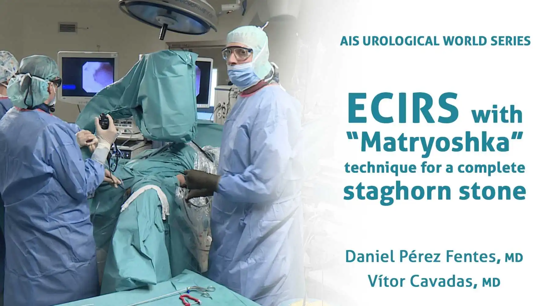
The patient was placed in the Galdakao-modified supine Valdivia position. A puncture was made under ultrasound and X-ray control in a posterior lower calyx, seeking a transpapillary puncture. The calyx entry was blocked, and the guidewire could not pass into the renal pelvis. An initial dilation of up to 16Ch was required to make room in this calyx access so to then place the guidewire in the ureter (“through and through” situation). A standard access (24Ch) was created using Amplatz serial dilators. A rigid nephroscope and lithotripsy using combined ultrasonic and ballistic energy with suction was used to remove the stone burden in the calyx access and renal pelvis. Flexible nephroscopy and laser were required to treat some fragments in the upper calyx and proximal ureter. Stones in the middle calyx were cleared out using a flexible ureteroscope inserted retrogradely (ECIRS) and extracted through the Amplatz sheath. A final endoscopic review did not reveal any residual fragments. At the end of the procedure a double J stent was placed without a nephrostomy tube, after having performed an endoscopic control of the percutaneous tract.
With Boston Scientific
Published
Jul 2019

Patient was placed in the Galdakao-modified supine Valdivia position. The double J stent was removed and a semirigid ureteroscopy was carried out immediately after. The ureteral stone was treated with Holmium laser and the fragments were actively removed using nitinol baskets. After having cleared out the ureter, a ureteral access sheath was inserted and a single-use flexible ureteroscope (LithoVueTM) was used to inspect the pelvicalyceal system. A percutaneous renal puncture was carried out under ultrasound and endovision control in a posterior lower calyx, looking for a transpapillary puncture. The tract was dilated up to 16Ch under endoscopic control. The different stones were treated using Holmium laser either through the rigid nephroscope and the flexible ureteroscope. Fragments were extracted through the Amplatz sheath, taking advantage of the “pass-the-ball” technique, with no residual fragments at the end. At the end of the procedure a double J stent was placed without nephrostomy tube, after having performed an endoscopic control of the percutaneous tract.
With Boston Scientific
Published
Apr 2019
Help your network discover Dr. Daniel Pérez-Fentes's clinical expertise.