

Innovation & Emerging Technology, Market Access & Commercialization, Clinical Outcomes & Patient Safety
Mission
To provide cost-effective and innovative surgical solutions to the worldwide healthcare community.
Vision
Continuous improvement of patient's health.
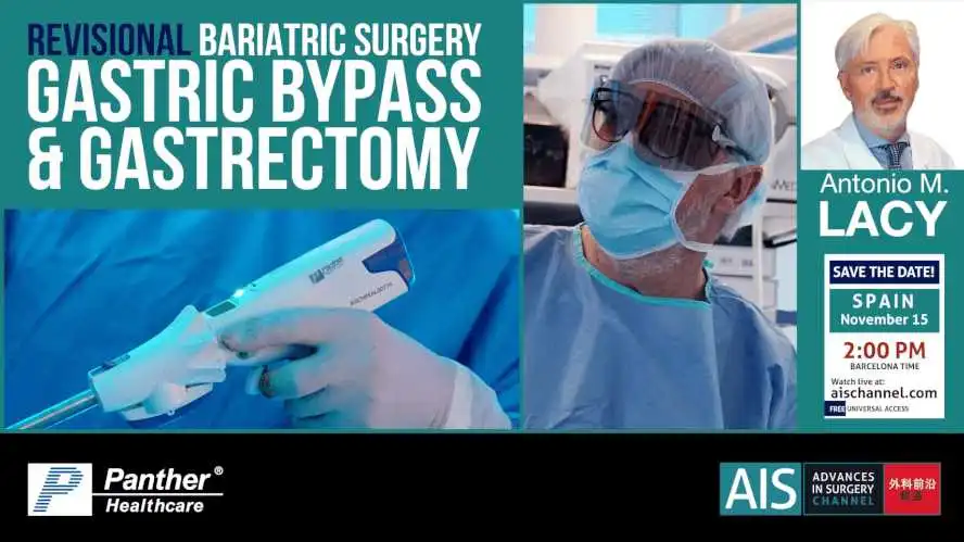
Dr. Antonio M. Lacy performs a Gastric Bypass to a 77-year-old woman with a medical history of hypertension under pharmacological treatment, currently untreated PSORIASIS ARTHRITIS, ARTHROSIS, episodic ASTHMA and VENOUS INSUFFICIENCY. Case record Gastroscopy: Tortuous esophagus without mucosal lesions. Peristalsis preserved. Cardia at 40cm from incisors. At subcardial level there is a "blind" cul-de-sac, about 3cm in diameter, with preserved gastric mucosa. Stomach: Gastric tubulization. Mucosa of the body and antrum with patchy areas of erythema, without erosions or ulcers. Normal pylorus. Duodenum without remarkable findings. BIOPSY - Mild chronic inactive gastritis with intestinal metaplasia without dysplasia. - Absence of glandular atrophy. - No microorganisms with morphology compatible with Helicobacter pylori were observed. Absence of malignancy. *Upper-GI series (barium swallow): Esophagus with tertiary contractions. Hiatal hernia. GER. Gastric tubulization. Good emptying of the stomach. Duodenum without alterations. *Manometry: Ineffective esophageal motility, according to the Chicago classification v.4. *IMPEDANCE - pHMETRY ESOPHAGEAL: Pathological acid reflux. DeMeester score 184.3. Conversion to Roux-en-Y gastric bypass Is proposed. Technique description Under general anesthesia, 12mm trocars umbilical, in epigastrium, right flank and left subcostal, 5mm Airseal trocars in left flank are placed. Careful dissection of previous gastric sleeve is performed. Postpyloric section is carried out with Panther® PEAL 60mm reinforced cartridge endostapler. Afterwards, hemostasis of duodenal stump is performed with clips. Creation of a 45mL gastric pouch using the same stapling device. Gastrectomy of the remaining stomach is completed. Dissection of the His’ angle and hiatus corroborating small (3cm) hernia. Posterior hiatoplasty is performed with interrupted 2/0 silk sutures. Section of the greater omentum. Section of the jejunum at 50cm from the Treitz angle. A 45mm lineal latero-lateral gastrojejunostomy is performed; closure of the gastro-enterotomy with a running 2/0 PDS suture. Section of the jejunal stump. Mechanical lineal latero-lateral jejuno-jeyunostomy is performed (alimentary limb 100cm); closure of enterotomy with running 3/0 PDS suture. Mesenteric and Petersen’s deffects closure with running 2/0 polypropilene sutures. Port-site revisión. Specimen removal through umbilical incision. Desufflation. Procedure steps Trocar placement Postpyloric section and hemostasis of duodenal stump Creation of gastric pouch Gastrectomy of the remaining stomach Dissection of the His' angle and posterior hiatoplasty Section of the greater omentum Gastrojejunostomy Jejunal stump section Jejuno-jejunostomy Mesenteric deffects closure Port-site revision Specimen removal Training Objectives To understand the surgical strategy for severe gastroesophageal reflux after sleeve gastrectomy. To identify the location and safe placement of trocars. Identification of the anatomical structures involved as well as the appropriate routes for dissection. Principles for the correct use of endo-staplers. Basis for the correct realization of an adequate gastric reservoir. Identify the basic maneuvers for the manipulation and measurement of bowel limbs. Identify and understand the fundamental steps and key maneuvers in the preparation of mechanical gastrojejunostomy and jejunojejunostomy. Recall the basics of intracorporeal suturing. Principles, justification and performance of the different closures of mesenteric defects. Systematic review of the abdominal cavity at the end of the procedure.
Published
Nov 2021
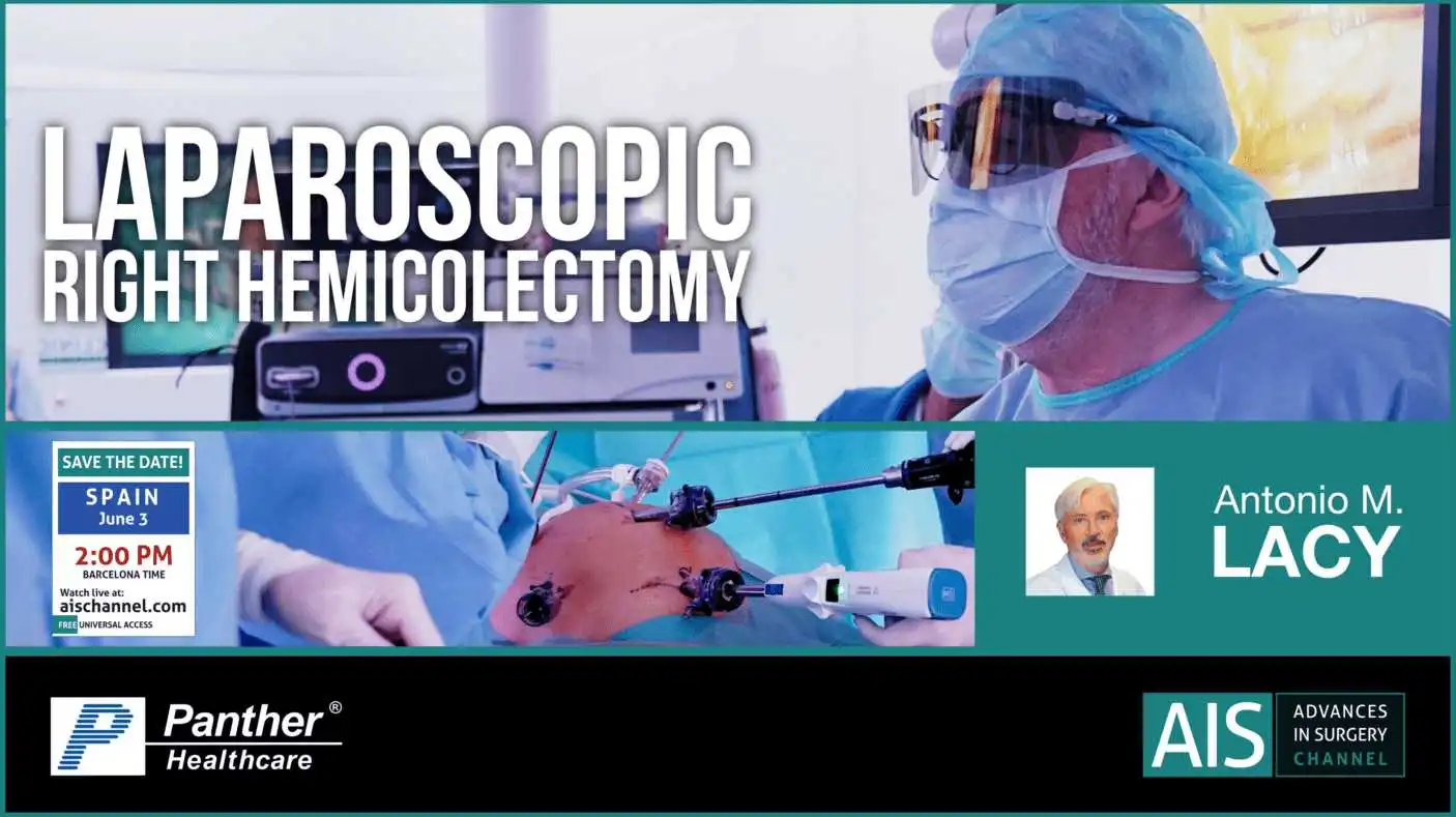
Dr. Antonio M. Lacy performs a laparoscopic right hemicolectomy to a 86-year-old male patient, diagnosed with an unresectable polyp of the cecum after diagnostic colonoscopy. Case Record: Male, 86 years old Unresectable polyp of the cecum with HGD Clinical History: 86-year-old male patient, diagnosed with an unresectable polyp of the cecum after diagnostic colonoscopy. Biopsy: high-grade dysplasia Proposed for pre-habilitation followed by conventional laparoscopic Right Colectomy. Other History: Medical History: Hypertension, Type 2 Diabetes, Atrial Fibrillation, Heart Failure, Stage III Chronic Kidney disease, Pulmonary dysfunction Surgical History: no previous surgeries Procedure Steps: Construction of the pneumoperitoneum with Veress needle Placement of an umbilical optical trocar followed by 4 working trocars: 5mm epigastrium and right upper and lower quadrants, 12 mm hypogastrium Inspection of the abdominal cavity Approach of the ileocolic vessels in a medial-to-lateral approach and ligation about 1cm from the origin Dissection of the posterior plane without disruption of the mesocolic fascia and leaving the duodenum and right ureter. Dissection of the mesocolon and mesentery of the terminal ileum Transection of the transverse colon and the terminal ileon and construction of an antiperistaltic latero-lateral stappled anastomosis after confirmation of vascularization with ICG. Trocar revision and extraction of the specimen. Learning Points: Dissection of the supplying vesselsDissection of the posterior plane without disruption of the mesocolic fasciaHow to perform an intracorporeal anastomosis
Published
Jun 2021
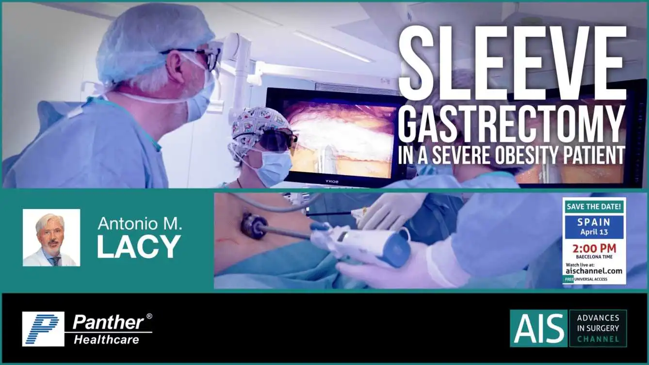
Dr. Antonio M. Lacy performs a Sleeve Gastrectomy in a Severe Obesity Patient Case Record: 44 y.o. patient diagnosed with morbid obesity. The patient has an BMI of 51 with no symptoms of gastroesophageal reflux disease. The patient was proposed for bariatric surgery and a sleeve gastrectomy was performed. Clinical History: 44 y.o. female patient diagnosed with morbid obesity, with a BMI of 51. Associated comorbidities: Type 2 Mellitus Diabetes treated with oral anti diabetic drugs and hypertension No clinical gastroesophageal reflux disease Proposed for a sleeve gastrectomy Procedure Steps: Patient in supine position, open arms and legs, reverse Trendelenburg Pneumoperitoneum creation Laparoscopic ports placement Marking of the inferior point of the dissection, 5 cm from the pylorus in the greater curvature Dissection of the omentum Revision of retro gastric surface Introduction of a 32 French bougie for gastrectomy calibration Transection the stomach with Panther 60 mm Stapling line coagulation control with hemo-clips Epiplon fixing to the remnant stomach Extraction of the removed stomach Closure of ports incisions Learning Points: Correct selection of the point for starting dissection Liberation of the His Angle Stapling line hemostatic control Selecting the correct endostapler
Published
Apr 2021
Share it with your network and help spread clinical knowledge.