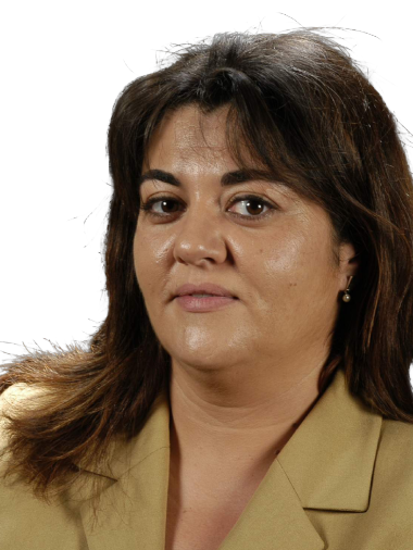
Dr. Salvadora Delgado completed her MIR training (1994-1998) and developed her professional career as a Gastrointestinal Surgeon at the Hospital Clínic de Barcelona. Since 2016, she has served as the Head of Service at HUMT, and since 2020, she has combined this role with the position of Director of the Surgical Care Department at HUMT.
She earned her Doctorate in Medicine from the University of Barcelona (UB) in 2001 and was later awarded the Extraordinary Doctorate Award in 2013. Since 2012, she has also been an associate professor at UB.
From the beginning of her professional career, Dr. Delgado has focused on advanced laparoscopic surgery, actively participating in the development of innovative surgical techniques for all digestive procedures (both upper and lower gastrointestinal tract), with a special interest in colorectal and bariatric surgery. Throughout her career, she has also performed surgeries in other general surgical areas, including biliary tract pathology, abdominal wall surgery, and splenectomies.
In the research field, Dr. Delgado has been an invited speaker at over 200 national and international conferences and has collaborated in organizing multiple courses and congresses.
She is a member of various surgical scientific societies and has served on the executive boards of some of them.
Selected publications from PubMed
Prostaglandin E2 Exerts Multiple Regulatory Actions on Human Obese Adipose Tissue Remodeling, Inflammation, Adaptive Thermogenesis and Lipolysis.
García-Alonso V, Titos E, Alcaraz-Quiles J, Rius B, Lopategi A, López-Vicario C, Jakobsson PJ, Delgado S, Lozano J, Clària J
PLoS One. 2016;11(4):e0153751 doi: 10.1371/journal.pone.0153751.
Accuracy of Vigileo/Flotrac monitoring system in morbidly obese patients.
Tejedor A, Rivas E, Ríos J, Arismendi E, Martinez-Palli G, Delgado S, Balust J
J Crit Care. 2015 Jun;30(3):562-6 doi: 10.1016/j.jcrc.2015.01.015.
Ventilation/Perfusion distribution abnormalities in morbidly obese subjects before and after bariatric surgery.
Rivas E, Arismendi E, Agustí A, Sanchez M, Delgado S, Gistau C, Wagner PD, Rodriguez-Roisin R
Chest. 2015 Apr;147(4):1127-1134 doi: 10.1378/chest.14-1749.
Hypertension remission 1 year after bariatric surgery: predictive factors.
Flores L, Vidal J, Canivell S, Delgado S, Lacy A, Esmatjes E
Surg Obes Relat Dis. 2014 Jul-Aug;10(4):661-5 doi: 10.1016/j.soard.2013.11.010.
Clinical markers of the hypercoagulable state by rotational thrombelastometry in obese patients submitted to bariatric surgery.
Taura P, Rivas E, Martinez-Palli G, Blasi A, Holguera JC, Balust J, Delgado S, Lacy AM
Surg Endosc. 2014 Feb;28(2):543-51 doi: 10.1007/s00464-013-3203-1.

When you have to chose who are the best patients to start performing the Cecil approach, everybody usually agrees in indicating the procedure for thin women with a middle rectal cancer. Nevertheless, older women have some specific characteristics that can make the procedure more difficult than expected, such as redundant colon, enterocele, lax tissues, etc. We can see how the combined approach will help us to increase the traction countertraction to solve the problems caused by laxity. Thanks to TaTME we were able define the anterior plan, which we would probably miss and follow too anteriorly using a conventional laparoscopic approach. After more than 200 cases, when people ask which kind of patient is the best to begin performing a trasanal total mesorrectal excision, we usually recommend a thin female with a middle rectal cancer. Thanks to their anatomical characteristics, women are the most appropriate patients as they have a wide pelvis, no prostate or seminal vesicles bordering on the anterior side, and provide the opportunity to mobilize the vagina for better definition of the surgical planes. But is a female pelvis always easiest? There are some characteristics, especially in older women, that can make this procedure challenging in this group of patients. In this video, we will try to show which difficulties you could find in a female pelvis. We present an 83-year-old woman with high blood pressure, dyslipidemia and Barrett’s esophagus. Due to rectal bleeding, the patient was diagnosed with rectal cancer 7 cm from the anal verge. Baseline staging revealed an mriT3N1 lesion plus resectable lung metastases by CT-Scan. This older lady received chemoradiotherapy with moderate response to a mriT2N0. Finally a low anterior resection with TME transanally and transabdominally was performed (Cecil approach). The patient was placed in a lithotomy position. Four trocars were used transabdominally, a 12 mm trocar was placed umbilically for the camera, and three 5 mm trocars were also placed, two in the right iliac fossa and one in the left hemiabdomen. A gel cap platform was used for the transanal approach. A wide pelvis, typical of a female patient, is visualized. Then the mesosigmoid peritoneum is incised to define the correct posterior plane. The inferior mesenteric artery is individualized. A high ligation of the artery was accomplihed by using the Ligasure™ after placing some proximal clips. The epiploic adhesions and Toldt’s fascia , which adhere the descending colon to the lateral abdominal wall, are resected, as well as the possible adhesions to the spleen. Simultaneously the transanal approach is performed. Firstly, after visualizing the rectal tumor, a pursestring suture is made with PDS or prolene to close the rectal lumen. After the rectum is closed a tattoo is made with the electrocautery. The down to up dissection is begun in a circumferential manner. We usually begin on the anterior side, where you can see some fibrosis secondary to the preoperative treatment. We continue afterwards on the posterior side preserving the mesorectum. And on the lateral side connecting the anterior and posterior dissections. At the same time, the abdominal dissection is continued into the pelvic space to perform a total mesorrectal excision. The left lateral side is also incised taking care not to damage the vessel and nerves. We continue our dissection through the presacral space preserving the mesorrectal fat. We can now see a deep Douglas down in the pelvis so we continue our dissection on the left lateral side, keeping the anterior side closed until the connection with the transanal team is made. This deep Douglas makes it difficult for us to maintain a correct traction countertraction so we require significant traction to proceed with the dissection and define the surgical plane. As you can see, the laxity of the tissues in older patients can complicate the procedure, making dissection more difficult. This patient has also a redundant sigmoid colon, which leads us to change the position of the instruments to maintain traction. Patient characteristics such as tissue laxity and medical situations such as enterocele can pose a real problem when finding the rectovaginal space. Thanks to the help of neumorectum and the work of the transanal team, we can find the correct anterior plane to incise the peritoneum more easily: not too close to the rectum where we could damage it or too close to the uterus or vagina, where we could find a bleeding plane. As you can see, the abdominal team is just increasing traction, pulling away the rectum from the vagina, in order to help the transanal team find the correct surgical plane. Coming back to the transanal approach, we will now focus on the anterior side, where the abdominal team is having problems defining the plane due to the laxity of the patient’s tissues. Thanks to the traction performed by the abdominal team, we can visualize the light and the correct plane through the anus. The rendezvous between both teams is achieved, thanks to the combined work. The correct plane is probably closer to the rectum than we had previously thought. Without the combined approach, we would probably have missed the anterior plane and done it too anteriorly. Once the correct plane is achieved, we can continue the abdominal dissection applying the right traction. With these lax tissues, traction for both teams is essential to perform lateral dissection with no injuries. The abdominal team retract from the distal side of the rectum to increase posterior traction, in order to complete the dissection transanally. In this way, the transanal team can perfectly see the correct posterior plane to finish the rectal resection and the specimen is introduced in the abdominal cavity. A prolene purse string on the distal rectum is made to perform the mechanical anastomosis. A silicon tube is connected to the spike of the stapler to exteriorize it through the anus more easily. The pelvis is now more narrow than we had thought at the beginning of the procedure, and the big uterus with an anterior myoma makes it even more difficult. Transanally, a clinch is introduced through the anus under laparoscopic supervision to exteriorize the proximal colon and the spike of the stapler to finish the anastomosis. The correct proximal colon position is checked. The colorrectal anastomosis is checked for leakage or bleeding and solved as soon as possible.
Published
Mar 2016

The patient is in a prone Trendelenburg position with legs wide open. A total of five trocars are used (a 12mm trocar in the supraumbilical position for a 30º camera, two trocars - 5 mm and 12 mm respectively- at the right iliac fossa, a 5 mm trocar at the left flank, and a final trocar at the epigastrium). The first step is the accommodation of the small bowel and then we start the dissection of the mesosigmoid. A key step during this maneuver is to perform traction of the mesosigma at the level of the inferior mesenteric vessels. The hook is used to open the peritoneum, and the pneumorectum helps by dividing the planes and improving visualization. Step by step, the inferior mesenteric artery is identified and sectioned using a LigaSure. The left ureter must be previously recognised. The surgery continues in a medial to lateral approach, stopping under the retroperitoneal plane. Now the inferior mesenteric vein is sectioned allowing better mobilization of the mesocolon. Now a window is opened in the mesocolon, and the most important landmark at this time is the pancreas and Riolan’s arcade. The assistant surgeon performs traction of the transverse colon towards the pelvis allowing good exposure of the omentum while the surgeon enters the lesser sac. Then the mesocolon is sectioned, and we continue to the retromesocolic space while respecting the arcade that runs along the top edge of the dissection. Finally we release the colon of the omentum thus completing the mobilization of the splenic flexure. In this case we followed a lateral to medial approach. The first step of the surgery was the same as in the previous patient. The hook was used to open the peritoneum and step by step the inferior mesenteric artery was identified and sectioned using a LigaSure™. We started lateral dissection using the hook. In this maneuver traction of the colon from lateral to medial by the surgeon and the assistant is very important. We must make a careful dissection without damaging the colon. Step by step we perform a left to right dissection releasing the colon and the mesocolon from the retroperitoneum above the pre-renal fat until the pancreas can be visualized. Finally we open the lesser sac and we release the omentum colon, thus completing splenic flexure mobilization following this approach. Sometimes use of a gauze in the retroperitoneal plane is useful to achieve better perception of the anatomy when following a medial to lateral approach. It also serves to assist in dissecting the correct plane. Then we complete the lateral dissection. Gauze serves as a guide to find the plane previously dissected from the medial area. In this way we combine both approaches. As in the previous cases we release the omentum colon, thus completing the splenic flexure mobilization.
Published
Feb 2016

In this video we will present some surgical scenarios showing the most common problems related with the pneumorectum that can arise when performing the Transanal TME. Transanal access platform set up: The first issue that may be found is that it is impossible to introduce the transanal access platform. This can happen because of a narrow or long anal canal, a hypertonic sphincter, or lack of relaxation. The use of a Lone Star retractor to expose the anus, good lubrication of the device and the use of dilators could decrease the situation. In addition, close interaction with the anesthesiologists is necessary, as this technique requires intense neuromuscular blockage to diminish sphincter pressure. Undesired effects of the pneumorrectum: Once the procedure has begun, we should take into account the effect of the transanal pneumorectum on the abdominal field. A retropneumoperitoneum can be generated, impairing the transabdominal team’s work. We strongly recommend that both teams work simultaneously (the Cecil approach). If only one team is available, the abdominal field should be performed prior to using the pneumorectum through the anus. The transanal pneumorectum can also insufflate the proximal colon, making the sigmoid resection difficult. To avoid this, the surgeon on the abdomen should clamp the distal sigmoid colon until the purse string is closed. If clamping the colon is not possible or has failed, the solution is deflating it using a rectal tube. Lack of rectal distention: Another common problem is the lack of correct distension of the rectal lumen when a pursestring has to be created or even during the dissection. The reason could be again incorrect neuromuscular blockage to avoid the effect of the surgical stimulus on the rectal wall and pelvic floor muscles. Another reason is high pressure from the abdomen. Try to reduce the CO2 on the abdomen by increasing pneumorectum pressure, until both sides are connected and pressure is equal again. Pursestring issues: Proper closure of the pursestring is essential to build a sealed cavity and achieve the exposure to work properly. A mistake during this step can create CO2 leakage in the rectal lumen giving rise to contamination and tumor spillage. Moreover, the proximal lumen will be insufflated which will hinder performance of the sigmoidectomy. To perform this step correctly we recommend making a tight knot, without the transanal access platform cap, ensuring a sealed cavity. If the error persists you should make a new suture or another stitch before moving on to the next step. Take care not to cut the suture due to too close rectal transection. Smoke evacuation: Another important issue to be concerned with is the kind of the device we use to provide the CO2 and evacuate the smoke. The conventional insufflator works in a pulsating manner, which increases rectum movement. In addition, every time we open a valve to evacuate the smoke, this modifies intraluminal pressure, resulting in significant pumping of the rectal tissue. All these factors hinder the procedure and tire out the surgeon so the procedure takes longer and the quality of the dissection may be compromised. Another type of insufflation device works by maintaining constant pressure on the pelvic cavity with a permanent smoke evacuation. Thanks to this device you can achieve a stable working field with a clear view. Which problems do you recognize on the next video? Exactly, we had problems with rectum distension as well as with smoke. What did we do? We achieved correct patient relaxation, low pressure in the abdominal cavity, and introduced CO2 insufflators that were able to maintain constant lumen cavity pressure. To sum up, with these easy tips and tricks you can solve some frequent problems that surgeons can find during the TaTME procedure.
Published
Feb 2016
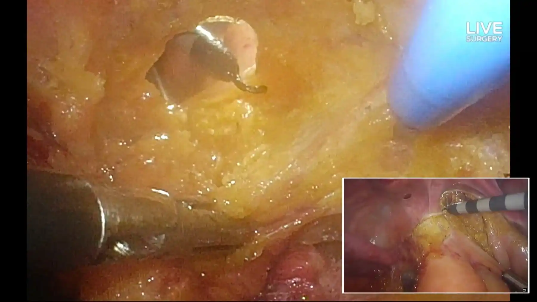
Performed by Antonio M. Lacy, Salvadora Delgado & María F. Hevia. Described by Joep Knol and Patricia Sylla in the Operating Room. Co-discussants: Richard J. Heald, Steven D. Wexner & H. Jaap Bonjer. Live connection during the surgery with Philip Quirke, Mariana Berho and Míriam Cuatrecasas for the pathology examination of the specimen.
With Medtronic
Published
Dec 2015

Case A 56-year-old female patient with no medical record. As a surgical history, she had a caesarean 28 years before. A study was initiated due to abdominal pain and altered bowel movements. A colonoscopy was performed, finding a stenosing rectal lesion 7 cm from the anal verge (pathology reported infiltrating adenocarcinoma, KRAS wild type). The study was completed with the following additional tests: Toraco abdominal Colono CT: which found a 1.2 cm subcapsular hypodense nodule at liver segment VII. The MRI confirmed that it was a metastasis. There were no other pathological findings. Rectal MRI: the tumor infiltrated the mesorectal fascia and the anterior peritoneal reflection and signs of extramural venous invasion. Uterus involvement was dubious (mri T4aN1) We also performed a transanal ultrasound and a PET scan which confirmed the diagnosis. Long course neoadjuvant treatment was given with 5 FU plus 45 Gy of RT. The re-staring MRI showed a slight decrease in the longitudinal diameter of the tumor, with persistent involvement of the mesorectal fat, the anterior peritoneal reflection and probably the uterus. This is a case of a stage IVa rectal cancer. As the liver mets required a minor liver resection, simultaneous surgery was attempted. Treatment The patient was placed in the Lloyd Davies position and a transanal access platform was placed. We introduced the GelPoint path transanally using 3 trocars (one for the 3D Olympus scope and two work trocars). A total of 4 trocars were placed in the abdomen: a 12mm port above the umbilicus for a 30○ scope. One 5mm port was placed at the right iliac fossa and a 5 mm port was placed on each flank. TRANSABDOMINAL PHASE: The patient was placed in Trendelenburg position, slightly to the right. First we made a silk stitch from the uterus to the abdominal wall, fastening it to improve exposure of the pelvis. Then we mobilized some of the sigmoid colon by means of sharp dissection using the hook. The left hand of the surgeons performs traction of the colon by pulling from an epiploic appendix and the right hand used the hook The bowel was accommodated to expose the mesosigma. The inferior mesenteric vessels were identified and dissected using the hook. It is important that the assistant surgeon maintain vessel traction to improve the visualization of the avascular planes. We observed the ureter before ligating the vessels for safety and to prevent unnoticed injuries. High tie arterial ligation was safely performed using a LigaSure™. The sigma was dissected, reaching the peritoneal reflection and leaving the colon free from the surrounding fat tissue. The descending colon was mobilized, from medial to lateral. Then we continued with the mobilization of the mesocolon to the rectum. The surgeon and its assistant should be synchronized to improve traction and exposure. At this point we realized that the cervix was affected, so it was decided to complete the surgery with a hysterectomy, bilateral salpingo oophorectomy. We used the LigaSure™ for this procedure. We completed the dissection of the uterus to perform an en bloc resection with the rectum. TRANSANAL PHASE: Meanwhile the transanal approach was under way. After the placement of the transanal device the pneumorectum was started, the tumor was identified and a ProleneⓇ pursetring was made setting the distal margin. The first step was to make the circumferential tattoo on the rectum wall. The dissection of the mesorectum began after sectioning the rectal wall. This maneuver must be performed perpendicularly to the rectal lumen and in a circumferential manner. The surgery progressed down-to-up while the abdominal team dissected the inferior mesenteric vessels and mobilized the sigmoid colon. The collaboration between the two teams is crucial at the highest part of the rectum. This helps to improve control of the specimen and prevents unexpected injuries. SIMULTANEOUS PHASE: Simultaneous work by both teams is important at this point, when they are working to improve exposure and clarify the planes in the same area. At the level of the anterior part of the rectum we had to cut the cervix to perform a block resection of the rectum and other structures affected. Finally we completed the surgery, removing the specimen. The specimen was extracted through a Pfannenstiel incision. An L-T stapled anastomosis was created under laparoscopic supervision with a 33 mm EEA. Finally a loop ileostomy was performed, ending the colorectal surgery. LIVER RESECTION: Then we performed liver surgery by a 3D laparoscopic approach. We placed another 4 trocars: 12mm ports on the right upper quadrant and the left flank, and 5 mm ports at the epigastrium and right subcostal. First we proceeded to section the falciform ligament and round ligament. Then we released the right hepatic lobe to right hepatic vein, and performed the section of the the right triangular ligament using the LigaSure. We dissected hepatic hilum to prepare it in case a Pringle maneuver was necessary. We used a ribbon to clamp the vascular hilum. Then we finally mobilized the right liver and held it using a liver retractor. Subcapsular hepatic metastases were identified in segment VII. The Pringle maneuver was performed for 11 minutes. Wedge resection of hepatic segment VII was performed leaving a sufficient free margin around the lesion. We used the Ligasure for the resection too. We extracted the specimen using an Endocath through the Pfannenstiel incision. We performed a cautious hemostasis of the liver using argon and perclot, ending the surgery. Outcome The surgery took 165 minutes. The patient started oral intake 24 hours after the surgery. During the postoperative period the patient presented with an urinary tract infection that was treated with an antibiotic. She did not present with fever. There were no other complications. She left the hospital on the 8th postoperative day. Pathological examination showed a pT4bN2a rectal adenocarcinoma. Hepatic resection was compatible with metastatic adenocarcinoma. Disease-free margins were found in both specimens. Two months later the patient is in good condition and receiving adjuvant therapy with FOLFOX.
Published
Nov 2015
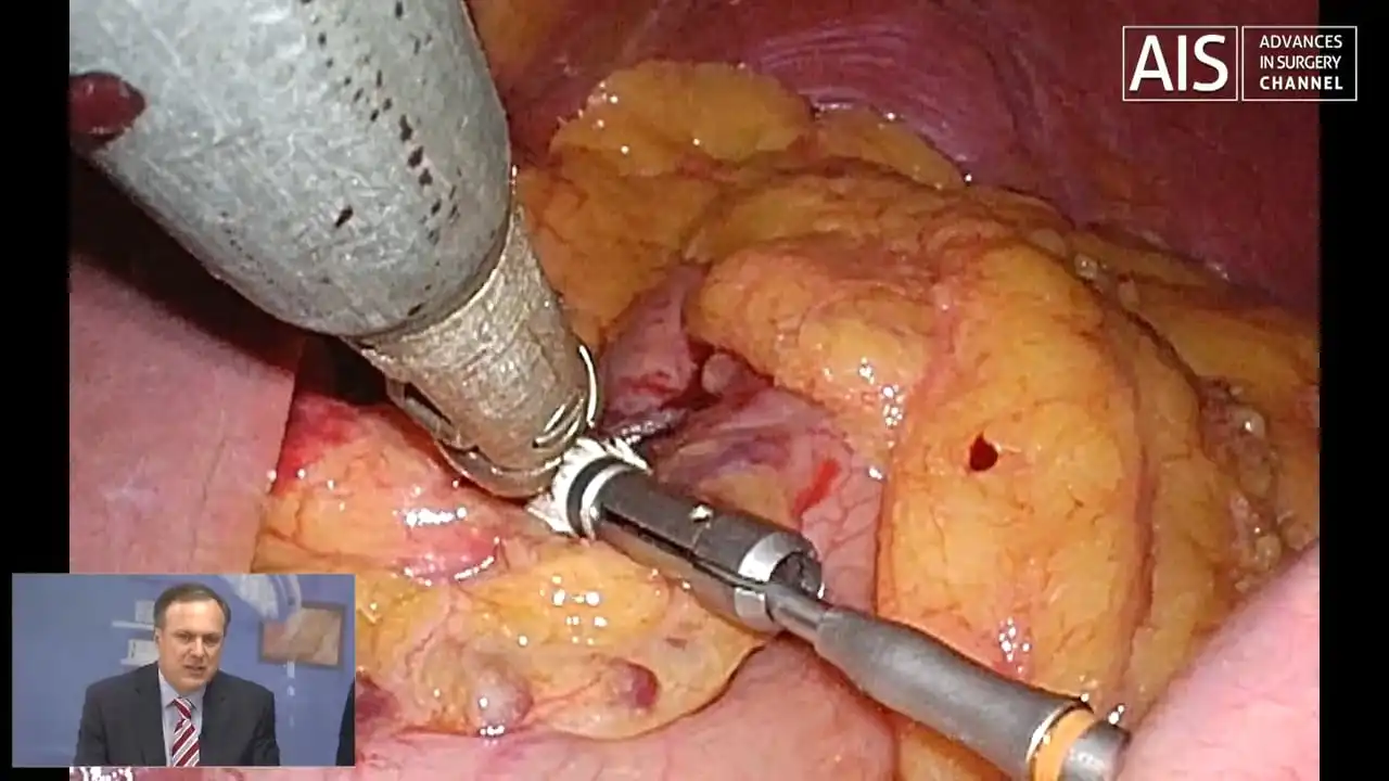
In this meeting, we will take a look at the Laparoscopic Roux-n-Y Gastric Bypass technique through its application in a live case of a morbid obese and diabetic patient, with live comments highlighting tips and potential pitfalls. A panel of recognised experts will then discuss the main hot topics regarding LRYGBP.
Published
Nov 2015
Help your network discover Dr. Salvadora Delgado's clinical expertise.