

General Surgery
General Surgeon, IQL, Spain
Dr. Raul Almenara is a general surgeon, member of the IQL team in Barcelona. He has an extensive professional career with stays in various surgical services among which includes the prestigious Cleveland Clinic in the United States. which in 2017 ranked 2nd in the ranking of the best hospitals in the United States. Member No. 14.791.
He is a specialist in colorectal surgery, gastrointestinal and hepato-bilio-pancreatic surgery as well as pelvic floor surgery.
Selected publications from PubMed
Time Interval Between the End of Neoadjuvant Therapy and Elective Resection of Locally Advanced Rectal Cancer in the CRONOS Study.
Guzmán Y, Ríos J, Paredes J, Domínguez P, Maurel J, González-Abós C, Otero-Piñeiro A, Almenara R, Ladra M, Prada B, Pascual M, Guerrero MA, García-Granero Á, Fernández L, Ochogavia-Seguí A, Gamundi-Cuesta M, González-Argente FX, Pons LV, Centeno A, Arrayás Á, de Miguel A, Gil-Gómez E, Gómez B, Martínez JG, Lacy AM, de Lacy FB
JAMA Surg. 2023 Sep 1;158(9):910-919 doi: 10.1001/jamasurg.2023.2521.
Functional Outcomes and Quality of Life After Transanal Total Mesorectal Excision for Rectal Cancer: A Prospective Observational Study.
de Lacy FB, Turrado-Rodriguez V, Torroella A, van Laarhoven J, Otero-Piñeiro A, Almenara R, Lacima G, Castells A, Lacy AM
Dis Colon Rectum. 2022 Jan 1;65(1):46-54 doi: 10.1097/DCR.0000000000001939.

Rectal cancer is the third most commonly diagnosed cancer in males and the second in females, only after lung, prostate and breast cancer. In 2018, 1.8 million new cases of colorectal cancer were diagnosed, while rectal cancer comprised about 700,000 new cases, the 8th cancer in terms of incidence. 310,000 deaths in 2018 make it the 10th most deathly. Only in Spain, rectal cancer claimed more than 4 thousand lives. The 5-year survival rate for rectal cancer is currently more than 75, with a 35% risk for metastatic diseases and local recurrence as low as 10-5%. It can be classified into clinical features like: Age: advanced ages are not associated with a poor prognosis. Performance status. CEA: the best tumor marker available to be used as an independent prognostic factor and as a monitor for recurrence of disease after primary tumor resection. Clinical stage: determined by TNM.Comorbidities. Histology: some variants, such as signet ring and mucinous, are associated with a worse prognosis, and also with a poor response to neoadjuvant treatment. Peritoneal involvement: associated with a decreased overall survival. Molecular markers: any genetic and epigenetic alterations that negatively affect the process of regulation, control of cell proliferation and differentiation, apoptosis, and angiogenesis. Neoadjuvant chemoradiotherapy (nCRT) + total mesorectal excision (TME) is considered nowadays the standard treatment for locally advanced rectal cancer. Since TME was described by Heald in the 80s, local recurrence has decreased from 30% to 10-5%, but still some advanced tumors had an adverse prognosis. That’s why some therapies like CRT are implemented. When we refer to neoadjuvant therapy (NAT), we’re practically talking about radiotherapy and chemo is administered as a radiosensitizer. Some countries advocate for classifying rectal tumors in categories (GOOD, BAD and UGLY), according to some characteristics which confer good or bad prognosis. According to this, patients are enrolled into 3 possible treatments: primary surgery, short course RT, long course RT. Summarizing, neoadjuvant therapy indications are: T4 tumors, N positive or with some adverse prognostic factors: Presence of extramural vascular invasion (EMVI) Suspicious mesorectal lymph nodes Circumferential resection margin <1mm Tumor height - Tumor in the anterior quadrant We deliver neoadjuvant therapy for the following reasons: Increasing downstaging Increasing the sphincter preserving procedures Increasing R0 resections Increasing the pathological complete response rate Increasing the pathological complete response rate: Short course radiotherapy, with immediate or delayed surgery Long course radiotherapy with conconcurrent chemotherapy and Total neoadjuvant treatment with both There’s currently an ongoing debate regarding the appropriate interval from the end of NAT and surgery that allows to achieve higher rates of pathological complete response (pCR). 8 weeks is considered standard, but interestingly some questions are yet to be answered: Do longer intervals increase the pCR? Is there any difference in DFS, OS or morbidity? The GRECCAR-6 study demonstrated that waiting for extended intervals does not increase the pathological complete response rate but increases technical difficulties during surgery and is associated with a higher complication rate. Probst et al on the contrary reflects that the key for obtaining a higher pathological complete response rate is increasing the interval from the end of NAT to surgery, without increasing the complications rate. It’s important to be aware of two concepts: Clinical complete response (cCR): no evidence of tumour when assessed by digital rectal examination (DRE), endoscopy-biopsy and MRI. Near Complete Response (nearCR): minor or equivocal findings during clinical examination which cannot be defined as cCR nor clinical bad response. We have several tools to evaluate the response to NAT: Endoscopy with a poor sensitivity of 50% and a negative predictive value of 11%, which can be explained by geographic misses when doing the biopsy. Digital rectal exam. MRI: has a special role, based on the tumor regression grade. PET TC: for the assessment of the metabolic response. Shows an accuracy of 91% which could be increased by a combination with clinical assessment. CEA levels. Endoscopy findings suggesting a pCR: Normal mucosa Whitening of the mucosa Telagiectasia Absence of tumor In reference to the MRI role, TRG referring to a classification of cancer response to preoperative treatment, can predict a prognosis of survival. It depends on what the radiologist sees in the images. It has its equivalent in the pathology report. TRG 1 and 2 are considered good response. Keon Jang et al in their meta-analysis evaluate the role of TRG. mrTRG1 for detecting pCR: Sensitivity: 32%. Specificity: 93%. They recommend using it as just ONE of the parameters for deciding to perform a less radical excision. Still more studies are needed, but the fact is that mrTRG is q0 times more likely to detect a pCR than proctoscopy/biopsy. Even though NAT is important for obtaining better oncological outcomes, it’s not harmless. This is demonstrated by the high rates of bowel dysfunction evidenced in patients treated by preoperative chemoRT, with various symptoms ranging from incontinence to anal blood or mucus loss, compared with those patients treated by surgery alone. Also they are more likely to develop some wound complications, hernias or bowel obstruction. Radical excision of the rectum entails a heavy burden for the patient: 28-40% of associated morbidity, 2% of mortality and also bowel, anorectal and sexual dysfunction. LARS or Low Anterior Resection Syndrome is defined as a disordered bowel function after rectal resection, leading to a detriment in quality of life. Comprises a bunch of symptoms like clustering of stools or fecal incontinence. Patients should expect some improvement over the first year, particularly in the first 6 months. The greater improvement in bowel function is in the first year and will never return to baseline. It’s worrying that 87% of patients are aware of their survival data, but only 53% were aware of functional alterations. POLARS, or the Pre-Operative Low Anterior Resection Syndrome score take into account 6 items for determining the risk of developing LARS: Age (older patients might have some functional bowel alterations that make them more likely to perceive the symptoms as minor), gender (more likely in women), Type of surgery (frequent in total mesorectal excision), Tumor Height (more in low and ultralow rectal tumors), Stoma (after reconstruction), and preop RADIOTHERAPY. The POLARS could help us to better inform patients and to decide the best treatment option for them. So, how to avoid morbidity? By selective use of radiation AND/OR increased adoption of nonoperative management and Watch & Wait. Pathological complete response (pCR) is seen in up to 20% after NAT. These patients could benefit from organ sparing management. There are a lot of studies trying to demonstrate the role of non-operative management in rectal cancer. Habr-Gamma, Garcia-Aguilar, Brown are obtaining great results with this strategy. Reported in the literature, watch and wait could obtain a 6% of local recurrence, 93% of disease free survival, 85% overall survival. There’s an international W&W database. The most striking result is the 24% local recurrence, knowing that these recurrences are mostly early recurrences (first 2 years), 97% endoluminal recurrences, and 88% of regrowths were treated with salvage surgery. On the other hand they reported 94% 5 year DSS. The follow up proposal during W&W is: 1rs/2nd year: DRE, Proctoscopy, CEA every 1-2 months + MRI/CT every 6 months. 3er-5th year: DRE, Proctoscopy, CEA every 6 months + MRI/CT annually. And if detection of relapse at any time, you should perform salvage surgery. The other way to avoid morbidity is the better selection of patients who received NAT. MERCURY and OCCUM are two studies that aim to demonstrate that delivery of Nat could be reduced so that we can reduce organ injury: MERCURY: evaluate primary surgery alone in MRI-predicted stage II or less and in MRI “good prognosis” stage III. 33% surgery alone, overall survival: 68%, Disease free survival: 85%, local recurrence: 3%.. OCCUM: reduce patients receiving NAT to 39.5%, found a Circumferential Resection Margin positive in 2.5% (surgery), 8.6% (NAT). R0: 99.2% (surgery), 96.8% (NAT). 5y-LRR and OS: awaited. Patients with locally advanced rectal cancer currently have a 30-35% risk of distant metastatic recurrence, which is the most frequent cause of cancer related death in this population. For solving this problem, the concept of TOTAL NEOADJUVANT THERAPY emerged. Consist in deliver chemotherapy as induction or consolidation concomitant with the conventional chemoRT. It has the following advantages: Improved delivery of planned therapy Increased downstaging Earlier introduction of optimal systemic chemotherapy Delivery of all chemotherapy preoperatively Reduce duration with a diverting ileostomy Based on two types of local recurrence, pelvic and in the lateral lymph nodes of the pelvis: the first associated with residual tumor cells in the pelvis and the second to lymph drainage. Pelvic recurrence is related to CRM & Histology and LPLN with CEA & swelling. Lateral pelvic lymph node dissection is perform in some Eastern countries like Japan. They demonstrated a 5-year relapse free survival similar in TME alone and TME plus LPLND (73.4% vs. 73.3%), explaining why they perform this procedure systematically. On the other hand it carries the risk of longer operation time and more complications. nCRT is recommended for avoiding pelvic recurrence and LPLND for LPLN recurrence.
Published
Feb 2020

Fecal incontinence is defined as the lack of control of defecation. It is a multifactor condition, involving such factors as mechanical disruption as well as neuromuscular or idiopathic dysfunction. Most of the time it is caused by a loss of anatomic continuity, for example after childbirth, surgery, or other traumatic injury. It frequently requires surgical intervention in an attempt to successfully restore normal physical structure. The causes are multiple, although its prevalence increases with age. After 50, prevalence rises to 11 % in men and 26% in women. The standard management for symptomatic fecal incontinence includes nonoperative management: - Pelvic floor exercises - Dietary changes - Surgery for repair sphincter defect We also know that 30% of fecal incontinence is linked to urinary incontinence. Sacral nerve stimulation has been approved for use in treating urinary incontinence in Europe since 1994, so it could also be a good option for these patients. Different studies have had good results showing that sacral nerve stimulation is a good option in patients with fecal incontinence, and is more effective than medical treatment alone. For all these reasons, the use of this therapy was proposed in patients who underwent rectal surgery and presented with Low Anterior Resection Syndrome (LARS). LARS is the major problem for rectal cancer patients after a low anterior resection. They have daily episodes of incontinence, obstructed defecation and constipation, causing a huge impact on the patients’ quality of life. 25% and 80 % of LARS patients develop symptoms following a sphincter-sparing rectal surgery. Mobilization of the rectum within the pelvis can lead to intramural nerve plexus injury, with long lasting sacral nerve terminal motor impairment. For individual patients, symptoms vary in type, severity, and duration. The risk factors for developing LARS are several, in particular: neoadjuvant radiotherapy, low anastomosis and anastomotic complications. There is evidence that denervation of the remnant sigmoid colon or left colon by means of operative maneuvers (e.g. ligation of a vascular pedicle) can result in a significant increase in motility. As a result, patients with LARS have a shorter colonic transit time and a greater increase in neorectal pressure after a meal compared to patients who do not have LARS. Removal of the rectum and in particular of the rectosigmoid junction eliminates the physiological distal control center for the regulation of bowel transit, leaving the bowel activities with no "brakes." This lack of distal negative feedback signals to oppose increased proximal colonic motility further exacerbates LARS symptoms. The sum of all these factors predisposes patients to develop LARS, which has a significant impact on their quality of life. The most frequently reported symptoms are: fecal incontinence, stool frequency, flatus incontinence, urgency, pad wearing, lifestyle modification, clustering and nocturnal incontinence. By means of the Wexner Score and the LARS Score we can classify this syndrome into minor and major according to the test score. Treatment in minor LARS ranges from dietary advice to loperamide, bulking agents and amitriptyline. In the case of major LARS the different options are stool training and advanced education, counselling, biofeedback, rectal irrigation and as a new option: Sacral nerve stimulation as a treatment for fecal incontinence due to LARS syndrome. Sacral nerve stimulation is a minimally invasive therapy. Different studies have shown its effectiveness in the treatment of pelvic floor dysfunctions, improving the severity of symptoms, restoring continence in patients and improving quality of life. The working mechanism is based on an effect on the autonomic nervous system, modulation of anorectal reflexes, modulation of the corticospinal pathway and changes in rectal sensitivity. It improves fecal continence by improving resting and squeeze pressures of the anal sphincter, as well as rectal sensation, together with stimulation on the external anal sphincter. Different studies show good results with this new technique in this type of patients, with improvement of symptoms in a high percentage of patients which also persists over time. We also see that quality of life improves, achieving a very significant decrease in the Wexner Score in different studies. The device system is an implantable programmable neuromodulation system that delivers electrical stimulation to the sacral nerve. It is designed to deliver therapeutic nerve stimulation through the following system components: a neurostimulator, a lead with programmable electrodes, and a lead extension. The system works by sending electrical pulses, which are produced by the neurostimulator, through the lead system. The implant goes through two phases. A first phase in which the device battery is external, checking if results are satisfactory. If they are, the final implantation is carried out in a second phase in which the battery is surgically implanted. For its placement it is necessary to select a location that meets the following conditions: - It is a minimum of 20 cm away from any other neurostimulator to minimize telemetry interference and possible inappropriate therapy. - It is on the opposite side of the body from another active implanted device (eg, pacemaker, defibrillator) to minimize possible interaction between the devices. - It is away from bony structures (e.g. 3 - 4 cm away) to minimize discomfort at the neurostimulator site. - It is away from areas of restriction or pressure to minimize the potential for skin erosion, patient discomfort, or damage to components. - It is in an area accessible to the patient for proper operation of a patient control device. - It is necessary to locate S3 for the needle implant. For this you have to identify middle line, identify sciatic notches, join the recesses and you will locate S3 is 2 cm from the midline. The needle should enter parallel to the midline and at the medial border of the foramen. If it is not placed in the correct position, electrode migration can occur causing the device to malfunction. Finally, we can say that with a good selection of patients, correct placement of the device and a good management program, sacral nerve stimulation can be successful in the treatment of these patients. In summary we know that, - LARS is related to a decreased rectal reservoir and denervation of the left colon during mobilization, which can lead to hypermotility of the neorectum and which may be a causal factor in the manifestation of multiple evacuations or urgency. - Authors have concluded that SNS is effective for all LARS symptoms. - Results of SNS implantation for fecal incontinence in LARS, showed significant decreases in fecal incontinent episodes, improvement in quality of life. - However, well-designed randomized controlled trials are needed to improve patient selection criteria for this procedure.
Published
Feb 2019

Clinical Case A 31-year-old female patient with a history of ulcerative colitis had undergone a total Proctocolectomy using a combined transabdominal and transanal approach (Cecil approach) to perform an ileoanal pouch. In addition, a loop ileostomy was made and subsequently closed after checking the anastomosis. Pathological examination showed a tubulovillous adenoma adenoma with low grade dysplasia on the rectal remnant close to the anastomosis, so periodic endoscopic controls were performed. However, during the last mucosal endoscopic resection, the finding was a tubulovillous adenoma with changes of adenocarcinoma and positive margins. Treatment It was decided to perform a resection of the anastomosis with mobilization of the ileoanal pouch through a combined abdominal and transanal approach. Once the transanal platform was introduced, the area of the anastomosis revealed an irregular mucosa. The rectal mucosa was tattooed with a cautery hook to ensure the distal margin of resection and guide the initial phase of the dissection. Subsequently, a careful dissection of the rectal mucosa was made, attempting to advance to the anastomosis in order to mobilize the pouch. We can observe that the tissue presents an important fibrosis due to previous surgery, which makes the procedure more difficult. During the anterior dissection, especial care was taken in order to avoid a vaginal injury. The dissection continued from distal to proximal, by means of the hook, going near the intestinal wall without injuring it. It is important to note that as in a total mesorectal excision, the dissection must be kept circumferential, from down to up, trying to always be working in the same plane. Once the area of the anastomosis was passed, it was important to be very careful not to injure the ileal pouch, even performing dissection with scissors when necessary. In case of hemorrhage we usually use bipolar, which also serves to dissect the different layers. Step by step, the dissection progressed towards the abdominal cavity. A good maneuver is the use of a gauze to push the specimen providing exposure and continue working on the dissection. At this point we can see how the adhesions are looser and are easily released with the scissors. In this phase of the surgery the abdominal team will help tractioning from the pouch to have tension and to favor the work from the transanal approach. Finally both teams joined their dissection planes. At this point, the transabdominal and the transanal teams can work simultaneously and assist each other to improve traction and identify the correct planes. With the assistance of both teams, the pouch dissection continued anteriorly and laterally towards the posterior side in order to completely release it. Once the pouch was released transanally, the mobilization of its proximal part was completed from the abdominal approach. At this point, the specimen is completely free and can be mobilized for transanal extraction, resection of the anastomosis area and preparation of a new coloanal anastomosis. The pouch is pulled distally by the transanal team to complete the procedure with a new ileoanal handsewn anastomosis without tension. In this case, an ileostomy was not performed. Outcome Operative time was 120 minutes. Patient resumed oral intake 8 hours after the surgery and was discharged on the 3th postoperative day. Pathological examination confirmed fibrosis without any signs of dysplasia or neoplasia. One month after surgery, the patient is in a good general condition and presents a correct anorectal functionalism.
Published
Aug 2017
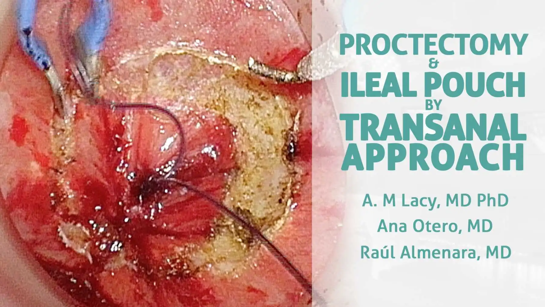
In selected cases of ulcerative colitis (UC), restorative proctocolectomy with a J-pouch and ileo-anal anastomosis (IPAA) is the treatment of choice. Total mesorectal excision has been preferred when performing the proctectomy, although a close rectal dissection (CRD), without following the "holy plane", has also been described. The transanal approach has evolved, and it is no longer limited to local excision of rectal neoplasms, with total mesorectal excision having currently become one of its main indications. Furthermore, this approach can also be used in cases of UC requiring surgery. Hybrid transanal CRD appears to be the next step in the evolution of minimally invasive transanal surgery.
With Olympus
Published
Jul 2017

Case A 59-year-old female patient with a previous history of diabetes mellitus, chronic bronchopathy, fibromyalgia, uterine fibroids, two vaginal deliveries and a surgical history of ovarian cyst resection. The patient had a complete rectal prolapse associated with moderate fecal incontinence. A normal colonoscopy, endoanal ultrasound, and defecography were recorded. A manometry was performed in which a moderate insufficiency of the internal and external anal sphincters was found. It was decided to perform a laparoscopic rectopexy. Treatment The patient was placed in the supine position with open legs. The surgeon stood at the right side of the patient with one assistant on each side. A total of 4 trocars were used. A 12mm trocar was placed in a supra umbilical position for a 30° scope. A 12 mm trocar was placed at the right iliac fossa and a 5 mm trocar was placed at the right flank, serving as working channels for the leading surgeon. One 5mm trocar was placed at the left flank as a helping channel for the assistant. First of all, the surgical field is prepared, leaving the pelvis free. To do this we separate the bowel and fix the uterus to the abdominal wall with a stitch. The patient is in the Trendelenburg position. Then we proceed to carefully open the peritoneum with the hook to dissect the mesocolon and release the promontory where the mesh will then be fixed. We continue with the release of the rectum at the level of the posterior side and towards the lateral sides, using the hook and ligasure. We progress distally by opening the peritoneal reflex at the level of the anterior side of the rectum. We dissect the anterior side of the rectum very carefully so as not to injure it, performing plenty of blunt dissection to avoid possible burns. At this level care must be taken not to injure the vagina. It is even helpful to perform a digital vaginal exam to make sure that we are not injuring it. Once we have completed the dissection we continue with the placement of the mesh. In this case we use a titanized mesh. It is important that the assistant maintain the traction of the colon to fix the mesh correctly. We make a total of four stitches at the level of the anterior side that we had released, two on each side, thus fixing the mesh to the rectal wall. Now we pull the mesh and fix it to the promontory that we released with tackers and one stitch of non-absorbable suture. Finally we close the peritoneum with a continuous bearded suture (V Lock) to cover the mesh and place the structures in their natural position. Outcomes The surgery took 90 minutes. The postoperative course was correct. The patient started oral intake 24 hours after the surgery and left hospital on the 2nd postoperative day.
Published
Jan 2017

Case A 72-year-old female patient with no medical history was diagnosed with an mriT3bN0 tumor 2.5 cm from the anal verge. The CT Scan ruled out any liver or lung metastasis. Long course neoadjuvant therapy was administered with good tolerance, and no toxicity was developed. The patient had a BMI of 16 Kg/m2 and an ASA score of II. Subsequently it was decided to perform an LAR by Cecil approach 12 weeks after treatment. Treatment The patient was placed in the Lloyd Davies position. A total of 4 ports were placed in the abdomen as well as a transanal access platform. The transabdominal team used a visualization system that combined HD and fluorescence imaging, while the transanal team used a 3D camera with a flexible tip as usual. The first step is to clamp the colon to work from the transanal approach. The bowel was accommodated to expose the mesosigma. After the placement of the transanal device the pneumorectum was started, the tumor was identified and the rectal wall was sectioned to make the pursestring and continue the dissection of the mesorectum. This maneuver must be performed perpendicularly to the rectal lumen and in a circumferential manner. In this case the transanal device was sectioned at the beginning of surgery as the tumor was very low. Subsequently both teams work simultaneously. The inferior mesenteric vessels were identified and dissected using the hook. High tie arterial ligation was safely performed using a LigaSure™. The descending colon was mobilized and then the sigma was dissected, reaching the peritoneal reflection and leaving the colon free from the surrounding fat tissue. Meanwhile the transanal approach was underway. The surgery progressed in down-to-up direction. Finally both fields were communicated completing the total mesorectal excision. Collaboration between both teams is crucial. The specimen was introduced into the abdominal cavity to improve traction until the resection was complete. The specimen was extracted transanally. At this point intravenous ICG pulses were administered to assess colon vascularization and to establish the suitable place to section the proximal margin. Given that a favorable response was not obtained after ICG administration it was decided to reintroduce the colon into the abdomen, partially improving its appearance, although not sufficiently. The colon was mobilized by decreasing the tension and traction of the mesocolon and ICG was administered again, now achieving a better response. Bad vascularization was objectified in the distal colon. It was therefore decided to mobilize the splenic flexure. The surgery continues in a medial to lateral approach, stopping under the retroperitoneal plane. We release the colon of the omentum to complete the mobilization of the splenic flexure. We also proceed to the section of the mesocolon preserving the left colic artery. We continue to section the mesocolon always keeping the pancreas and Riolan’s arcade in mind. Finally the entire colon was mobilized. It was decided to make an auxiliary Pfannenstiel incision to examine the colon and ensure a well-vascularised anastomosis with no tension. ICG was injected again into the exteriorized specimen, achieving a good result, clearly identifying the section point and checking good vascularization of the coloplasty. A T-T manual anastomosis was performed, completing the procedure. A loop ileostomy was built as the patient had received preoperative radiotherapy and the anastomosis was low in the pelvis. Outcome The surgery took 210 minutes. The patient started oral intake 24 hours after the surgery and left hospital on the 3rd postoperative day with no complications. We can say that ICG provides an option to evaluate the perfect site to perform an anastomosis in colorectal surgery and makes it possible to ensure good vascularization in a simple and safe way.
Published
Nov 2016
Help your network discover Dr. Raul Almenara's clinical expertise.
