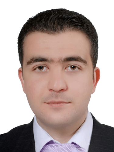
After he graduated from the medical school in 2012, he joined the Department of General Surgery at Damascus University as a Post Graduate Resident for a five-year residency program. He got his MS in General Surgery in June 2018. During that period, he operated and helped in hundreds of cases in both elective and emergency surgeries, including several blast injuries. In July 2018 he joined GEM hospital and research centre in Coimbatore-India as an International fellow, and he got a Diploma in Laparoscopy in September 2018. Additionally, he became a lifetime member of the Association of Minimal Access Surgeons of India. In September 2018, he led, in Syria, GlobalSurg3, an international prospective cohort study about Quality and outcomes in global cancer surgery. In July 2019 He was awarded Chevening Scholarship, the most prestigious UK scholarship, to study a master in Advanced Minimal Invasive Surgery at University College London. He is currently working as a specialist General Surgeon at Badr Al Sama group of hospitals in Oman. He has a particular interest in laparoscopic and robotic colorectal surgeries, and he looks for further training in that field. Dr ALShaar is the father of two boys, and he likes travel, cooking and classical Arabic music.

Surgical Management of Hemorrhoids · Surgical interventions are recommended in patients who have failed medical and office-based management. Usually, they are indicated for the higher grade of hemorrhoids (grades III-IV). · The most commonly used surgical interventions are the traditional excisional hemorrhoidectomy, including open (Milligan-Morgan procedure) and closed (Ferguson procedure) hemorrhoidectomy. · New surgical procedures such as stapled hemorrhoidectomy and hemorrhoidal artery ligation (HAL) were developed to lower the incidence of postoperative complications; however, they are criticised for high recurrence rates, high costs and unique complications. · Here we will describe each of these procedures briefly. Ferguson’s hemorrhoidectomy (closed technique) · It is the most commonly performed hemorrhoidal procedure in the United States. · Dr Lynn Ferguson first described it in the early 1950s. · Anaesthesia can range from a combination of local anaesthesia with intravenous sedation to full general anaesthesia. · The patient’s position can be lithotomy, prone jackknife, or left lateral decubitus. · Before starting the operation, Identifying the hemorrhoid complex by digital rectal examination and anoscopy is mandatory. · Adding epinephrine 1:200,000 to the local anaesthetic can help to decrease bleeding during the procedure. · Starting at the perineal margin an elliptical incision is made around the hemorrhoid, and a proportional incision should be made so that the length of the incision is approximately three to four times longer than its breadth. · The hemorrhoid is excised after it is elevated off the underlying sphincter muscular fibers and its vascular pedicle is clamped and ligated with an absorbable tissue at the apex. · The hemorrhoidal bundle is then liberated from the sphincter complex using either electrocautery or a bipolar sealing device, which was better in terms of reducing operation time and postoperative pain and bleeding. · Then the resultant hemorrhoidal bed is approximated using absorbable suture, and usually, a small opening is typically left distally to improve cosmesis and facilitate and drainage of any retained fluid in the hemorrhoidal bed, reducing the risk of perineal sepsis. · During excision multiple hemorrhoids, it is mandatory to maintain adequate skin and tissue bridges between the excision sites to minimise the risk of postoperative anal stenosis. Milligan-Morgan hemorrhoidectomy (open technique) · It is the most commonly used in the United Kingdom, originally described by Milligan and associates in 1937. · A V-shaped incision is made in the anoderm that does not extend into the mucosa. · The pedicle created by traction on the artery forceps then removed from the longitudinal strands of fascia and muscle of the internal anal sphincter. · Dissection can be achieved with scissors or more usual monopolar diathermy. As the dissection proceeds upwards, the underlying mucosa must be divided on each side of the pedicle. · The mucosal incisions should converge towards the apex of the pedicle in order to avoid leaving a broad, bulky mucosal pedicle. · The apex of the pedicle is then transfixed with 0 or 1/0 Vicryl, and the mucosal aspect of the hemorrhoid then removed. · Mucosal bridges of approximately 1 cm should be maintained between each hemorrhoid to avoid causing anal stenosis. · After the internal and external components of hemorrhoids are excised, a three-leaf clover pattern remains and is allowed to heal by secondary intention. · The technique requires careful attention to avoid damage to sphincter muscles. This technique has the advantages of decreasing operative time and postoperative pain; however, it typically takes longer healing time compared with the closed technique. Excisional hemorrhoidectomy has excellent results, minimal recurrence rates, and few complications yet are associated with significant postoperative pain. Stapled hemorrhoidectomy · It is an endo-stapling technique employed to deal with prolapsing hemorrhoids or mucosal prolapse, First described in 1993 by Longo. · It reduces the mucosal and hemorrhoidal prolapse by excision of a transverse circular band of the prolapsed anal mucosa between the distal rectal ampulla and the proximal anal canal aiming to restore the normal topographic relationship between the anal mucosa and the anal sphincters. · The technique is usually performed under general anaesthesia, and the patient is usually positioned in the prone jackknife position or in the lithotomy position. · With the anoscope in place, a pursestring suture is placed circumferentially into the submucosa approximately 2 cm above the transitional zone. · The head of the stapler (similar to an EEA, but the head is not detachable) is then introduced into the rectum past the pursestring suture. · The pursestring is tied down around the stapler, and then the anvil is very slowly closed while giving gentle traction on the pursestring suture externally. · Once closed, the stapler is fired and then removed along with the excised tissue. · The staple line should be inspected carefully for bleeding and may require suture ligation. When compared to excisional hemorrhoidectomy, this technique showed less postoperative pain, shorter inpatient stay and quicker return to normal activities, but the recurrence rates were higher. Additionally, unique complications were reported such as persistent anal pain, rectal perforation, rectal obstruction, rectovaginal fistula and retroperitoneal sepsis. hemorrhoidal Artery Ligation (HAL) · This procedure is an evolution of an older technique revived by Farag in 1978, who advocated suture of what he described as the perforating veins at the base of the vascular cushion. · The key principle of these procedures involves the ligation of terminal hemorrhoidal branches of the superior hemorrhoidal artery after accurate localisation using the Doppler flowmeter. · Morinaga et al. seem to have been the first to use the technique in 1995; they devised a proctoscope that incorporated within it a Doppler transducer, which they named the Moricorn, and through which the hemorrhoidal artery (as opposed to the vein) could be identified and sutured. · This specialised anoscope with a Doppler is inserted 4–6 cm from the anal verge and above the dentate line. · The Doppler is used as the anoscope is rotated until one of the feeding arteries is identified and absorbable figure 8 knot suture is ligated close to the probe sensor at a depth no greater than 8 mm · The anoscope is rotated until all of the significant arteries are identified and ligated. · A second round of ligation after the withdrawal of the probe handle 1–1.5 cm can also be performed. · Recto-anal repair (Rectopexy) can be added at the same setting to minimise prolapse by running stitch applied to each prolapsing hemorrhoid, suturing from the proximal to distal and no closer than 5–10 mm above the dentate line to avoid pain. References 1· The ASCRS Manual of Colon and Rectal Surgery, Third Edition 2019 2· Keighley & Williams’ Surgery of the Anus, Rectum and Colon. Volume 1, 2019. 3· CURRENT SURGICAL THERAPY, 13 Edition, 2020.
Published
Nov 2020

Five things you should know about the Surgical Anatomy of the Thyroid Gland The thyroid gland is located in front of the neck below the thyroid cartilage. The average weight of the thyroid gland, in healthy adults, is 15–30 g. It consists of two lobes connected by an isthmus in the middle. The pyramidal lobe, which ascends from the isthmus or the adjacent part of either lobe up to the hyoid bone, can be seen in 20 % of the patients. Arterial blood supply of the Thyroid gland The main arterial blood supplies of the thyroid gland come from: Superior Thyroid Artery The first branch of the external carotid artery, it enters the upper pole of the thyroid on its anterosuperior surface. Inferior Thyroid Artery Usually arises from the thyrocervical trunk upward in front of the vertebral artery and Longus Colli to the lower pole of the thyroid gland. Before entering the thyroid, the artery usually divides into 2–3 branches. The inferior thyroid artery and its terminal branches are closely associated with the recurrent laryngeal nerve at the insertion of the thyroid gland, and innumerable variations have been described, so care must be taken in all cases to avoid injuring the nerve. Thyroid IMA Artery An unpaired artery in the anterior surface of the trachea, which can be seen in 3–10 % of patients. It usually arises from the brachiocephalic trunk (innominate artery), and occasionally it may arise from the aorta, the right common carotid, the subclavian, or the internal thoracic artery. It supplies the lower part of the thyroid gland. Venous drainage of the Thyroid gland: A well-developed thyroid venous plexus usually drains through the inferior thyroid vein to the left brachiocephalic (innominate) vein. The superior and middle thyroid veins drain to the internal jugular vein. Nerves must be aware of during thyroidectomy During thyroidectomy, Care must be taken to preserve the right and left recurrent laryngeal nerves in addition to the superior laryngeal nerves. The left recurrent laryngeal nerve branches from the vagus nerve, and loops under the arch of the aorta. It usually ascends along the tracheoesophageal groove with a straight course. The right recurrent laryngeal nerve loops around the right subclavian artery. It courses more obliquely and more lateral than left. The superior laryngeal nerve, which is a branch of the vagus nerve, takes part in the pitch of the voice. It descends, by the side of the pharynx, behind the internal carotid artery and divides into two branches: external laryngeal nerve as a motor nerve and internal laryngeal nerve as a sensory nerve. There is a close relationship between the superior thyroid artery and the external branch of the superior laryngeal nerve; therefore, it is recommended to ligate the superior thyroid arteries as low as possible on the thyroid gland to avoid injuring the external branch of the superior laryngeal nerve. Parathyroid Glands Usually, there are four parathyroid glands, which are located on the rear surface of the thyroid gland. The regular size of the parathyroid gland is less than 1 cm and weighs 25–40 mg. They are yellowish-brown and may be distinguishable from the thyroid gland. The superior parathyroid glands are normally located on the dorsal aspect of the upper pole of the thyroid gland. The inferior parathyroid glands are usually located posteriorly to the lower pole of the thyroid but are commonly found within the thymus in the neck or upper mediastinum. The inferior parathyroid gland is anterior and medial to the recurrent laryngeal nerve, while the superior parathyroid gland is located posterior to this plane. The principal arterial blood supply to both superior and inferior parathyroid glands is described as the inferior thyroid artery; there are also other blood supplies from the superior thyroid artery or anastomotic vessels of superior and inferior thyroid arteries. Lymphatic drainage of the Thyroid gland In the first place, immediate lymphatic drainage is to the periglandular nodes. And it courses to the prelaryngeal (Delphian), pretracheal, and paratracheal lymph nodes. These lymph node groups are called central lymph nodes or central neck compartment; (levels IA, IB, and VI), and they are the most common site of metastasis from thyroid cancer. The central neck compartment boundaries are: the hyoid bone superiorly, the suprasternal notch inferiorly, and the medial border of common carotid artery laterally. The second part of lymphatic drainage is the lateral neck region, levels IIA, IIB, III, IV, VA, VB. References 1. Youn Y-K, Lee KE, Choi JY. Surgical Anatomy of the Thyroid Gland. In: Color Atlas of Thyroid Surgery. Berlin, Heidelberg: Springer Berlin Heidelberg; 2014:1-10. doi:10.1007/978-3-642-37262-9_1
Published
May 2020
Help your network discover Dr. Muhammad Alshaar's clinical expertise.