Selected publications from PubMed
CRISPR/dCAS9-mediated DNA demethylation screen identifies functional epigenetic determinants of colorectal cancer.
Tejedor JR, Peñarroya A, Gancedo-Verdejo J, Santamarina-Ojeda P, Pérez RF, López-Tamargo S, Díez-Borge A, Alba-Linares JJ, González-Del-Rey N, Urdinguio RG, Mangas C, Roberti A, López V, Morales-Ruiz T, Ariza RR, Roldán-Arjona T, Meijón M, Valledor L, Cañal MJ, Fernández-Martínez D, Fernández-Hevia M, Jiménez-Fonseca P, García-Flórez LJ, Fernández AF, Fraga MF
Clin Epigenetics. 2023 Aug 24;15(1):133 doi: 10.1186/s13148-023-01546-1.
Complete Mesocolic Excision and D3 Lymphadenectomy versus Conventional Colectomy for Colon Cancer: A Systematic Review and Meta-Analysis.
Díaz-Vico T, Fernández-Hevia M, Suárez-Sánchez A, García-Gutiérrez C, Mihic-Góngora L, Fernández-Martínez D, Álvarez-Pérez JA, Otero-Díez JL, Granero-Trancón JE, García-Flórez LJ
Ann Surg Oncol. 2021 Dec;28(13):8823-8837 doi: 10.1245/s10434-021-10186-9.
Transanal total mesorectal excision: surgical technique description and outcomes.
Trépanier JS, Fernandez-Hevia M, Lacy AM
Minim Invasive Ther Allied Technol. 2016 Oct;25(5):234-40 doi: 10.1080/13645706.2016.1199434.

One of the main goals of AIS Channel is to identify and broadcast technological innovations that improve surgical outcomes. This year, our summer event was held on July 14th: a live streaming congress that focused on oncologic assessment with fluorescence imaging. Steven D Wexner designed a robust agenda that included the most relevant data aimed at enhancing lymph node assessment prior to, during and after surgery. This will potentially lead to a more precise and tailored treatment. Experts from the United States, the United Kingdom, Spain, Ireland and Switzerland brought the latest data, exchanged ideas, and answered the questions sent by viewers from all over the world, providing a global opinion. Simultaneous Live Surgeries were performed by Antonio Lacy, Raquel Bravo & María Fernández-Hevia, using the laparoscopic and robotic approach for treatment of rectal cancer. This was an exciting experience moderated by David Jayne. During the procedures, several ICG guided strategies were used for the lymphovascular dissection, tissue assessment, and anastomotic perfusion. Both resected specimens were evaluated by Mariana Berho, who confirmed a correct dissection. Finally all the experts gave their final statements and closing remarks which you may see in this video. The 2016 AIS Summer Event was once again a huge success in terms of audience. Thousands and thousands of colorectal surgeons from 99 countries logged in and watched the entire event live. Once again, AIS Channel is consolidating itself as the most innovative and popular digital tool in surgical education, through its approach of providing the entire global medical community with the best training.
Published
Aug 2016

Ureteral damage is one of the intraoperative complications most feared by surgeons. The first thing that we must all have in mind is avoiding the section of such an important structure. There are two possibilities regarding prevention. Some surgeons prefer to perform a complete dissection of the inferior mesenteric vessels and the retrocolic space which allow them to look for the ureter before the vessel section and complete the procedure. On the other hand, there are surgeons who argue that if you are in the correct surgical plane, preserving correctly the retrooperitoneum, there is no need to look for the ureter before continuing the procedure. In our group we are in favor of the latter. Nevertheless there are some dificult situations such as obese patients, hostile abdomen with a lot of adherences, fatty mesocolon, dolicosigmas, etc. which increase the risk of ureteral lesions. But this unpleasant complication is not a synonymous with conversion to open surgery. In experienced hands, the laparoscopic approach, together with the use 3D view systems, provides a high definition view which makes it possible to diagnose complications, guide an ureteral catheter correctly and suture the ureter over it. In the next video we will show a ureteral section that took place during the abdominal access in a transabdominal-transanal total mesorrectal excision. We describe the main surgical steps to complete its correct repair. Case The clinical case is a 77-year-old woman with a BMI of 34 Kg/m2 and a diagnosis of middle rectum cancer cT3N0 treated with neoadjuvant chemoradiotherapy. Due to obesity and intrabdominal adherences, the definition of the surgical planes was especially difficult and a ureteral lesion occurred. Treatment The perianal approach performed during the TaTME allowed accurate pelvic dissection. The increased traction obtained by the two teams working at the same time better defines the surgical plane and improves the visualization of the surrounding organs. These advantages are especially important in both laterals and the anterolateral region to preserve the nerves, the vessels and the entrance of the ureter into the pelvis in the upper rectum. The anatomical landmarks from the pelvis should be known. After the resection the correct intact presacral plane could be visualized as well as the hypogastric vessels and nerves. Thanks to this close evaluation we were able to observe a lateral damage of the ureter with a partial section. In the video you can see the ureter, with its creeping movement, crossing over the division of the iliac artery. This unpleasant complication is not synonymous with conversion into open surgery. In experienced hands, the laparoscopic approach provides a high definition view which makes it possible to diagnose the complications, guide a ureteral catheter correctly and suture the ureter over it. The transanal approach combined with a 3D camera provided a direct view of the ureter, so we were able to make a first simple prolene stitch through the sectioned ureter. To check the correct lumen of the ureter we introduce an avocath, also transanally. You can introduce the catheter through the 10 mm trocar or taking out the silicon device, which is also easy and fast. The other surgeon on the abdominal side can help by performing gentle traction of the proximal side of the ureter. This way we can check that we have properly respected the lumen of the ureter before finishing the end to end anastomosis with 3 single stitches. Finally, a pig tail catheter is introduced by cystoscopy localizing the left ureteral lumen. Outcome The patient’s evolution was correct, and she was discharged 6 days after surgery. In conclusion, the ureteral injury is a feared intraoperative complication that can be solved laparoscopically. Its transanal repair is feasible and safe, providing a direct high definition view of the damage. The 3D view can add depth, helping us to perform such a demanding procedure.
Published
Jul 2016
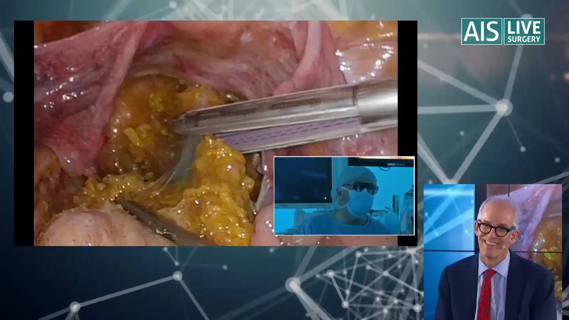
AIS Channel broadcast two simultaneous LIVE Robotic and Laparoscopic Surgeries performed by Antonio M. Lacy, Raquel Bravo, Maria Fernandez-Hevia and Ana Otero. David Jayne commented the procedure inside both Operating Rooms with Steven D. Wexner in the Optimus Room.
Published
Jul 2016

Case The patient was a 72-year-old female with high blood pressure, dyslipidemia and rheumatoid arthritis who was diagnosed with a well differentiated mriT1-N0 adenocarcinoma 7cm from the anal verge. Clinical staging by CT-Scan ruled out distant metastasis. Local resection was performed through the TAMIS approach and the pathology analysis revealed an pT3 high grade mucinous adenocarcinoma with a lymphatic invasion, low grade budding, lateral free margins and a deep margin at 1.5mm. The options presented were the following: Watch-and-wait approach Chemoradiotherapy and surveillance Chemoradiotherapy and radical surgery Radical surgery without neoadjuvant therapy Treatment Our final decision was taken in a multidisciplinary committee involving oncologists, radiologists, gastroenterologists and surgeons. The option selected was radical surgery with no neoadjuvant therapy (which was also the most voted option in our survey). To continue, we will show the complete surgery, which was performed laparoscopically: a low anterior resection through the Cecil approach. The patient was placed in the supine position with open legs. A total of 4 trocars were used. A 12mm trocar was placed in the umbilical position for a 30° scope, and three 5 mm trocars served as working channels on the right iliac fossa, and on each flank. This video focuses on the transanal approach with some reference to the abdominal approach. Once the transanal device was introduced, we were able to explore the rectum. You can see the perfectly closed scar of the resected tumor 6 weeks ago. Since the height of the scar was very low, we decided to perform the section of the rectum first, in order to have sufficient oncologic margin and to close the rectum safely. A tattoo is marked circumferentially to guide the opening of the rectal wall (first the mucosa and then the muscular layer).Step by step we opened all layers of the rectum to achieve a complete circumferential section. In this case, as the patient was a woman, we took special care on the anterior side as we could injure the vagina. In a man we would have to avoid injuring the prostate at this level. The hook is used to open the rectum and the pneumo helps to expose the avascular plane. During this step the colon is clamped from the abdominal approach to prevent it from being filled with CO2. Once the section of the rectum was performed, a purse-string was made to close the rectal lumen. The pursestring should be closed tight to avoid pneumo leakage. Then we introduced a gauze to help with handling and continued with the dissection of mesorectum. The plane must be followed from where it is identified to the point where the dissection must continue. Mobilization must be circumferential, imagining a cylinder inside the pelvis curve. Avoid getting deep into one plane, as the rectum will retract and the quality of the dissection will be compromised. In the posterior plane, the presacral fascia can be damaged and there may be bleeding from the presacral vessels. The bipolar is sometimes a useful tool to control the situation. We continued with the lateral dissection, trying not to create any holes and stay always within the same plane circumferentially. Finally we connected both approaches. Combined work from the transanal and transabdominal teams saves time and improves the dissection, helping to clarify some hard-to-reach planes. The dissection must be symmetrical. Gradually we see how the resection is completed by combining the work of two teams at a time, and is both safe and reliable. Finally, we extracted the piece through the anus aided by the abdominal team, ensuring that the colon had no tension and was in the correct position to perform the anastomosis. Coloanal manual side to end anastomosis was performed. First of all, the long star was placed and we made four cardinal vicryl stitches in the distal rectum. Then the rectum was exteriorized and cut with an endoGIA. The colon was opened laterally so as to perform a side-to-end anastomosis. The previous sutures made were completed with four stitches in the proximal colon after opening the colonic wall. The anastomosis was completed with single vicryl stitches between the previous cardinal sutures. We can check that we have a correct colonic lumen during the procedure. We finally checked hemostasis and completed the surgery. Outcome The surgery took 100 minutes. The patient started oral intake 48 hours after the surgery and left hospital on the 5th postoperative day. Pathological examination ruled out scar fibrosis and malignancy in the prior resection. The mesorectum was complete. Metastasis was observed in one of nine lymph nodes found.
Published
Jul 2016

There is no doubt that the total mesorectal excision (TME) is the best surgical treatment for rectal cancer. However it may be a challenging procedure with complications between 30 - 68%. That is why among other reasons radical surgery should be performed in patients with a favorable balance between risk and benefits. There is a group of patients with early rectal cancer (cT1 sm2/sm2 cN0) that may be treated with local resection, achieving curative surgical treatment with less complications. In this lecture María Fernandez-Hévia from the gastrointestinal surgery department at Hospital Clinic of Barcelona will explain the modern surgical treatment tailored to patients and tumor characteristics.
Published
Jun 2016

On April 21 2016, AIS Channel broadcast a live surgery in which Antonio Lacy, Ricard Corcelles and Maria Fernandez-Hevia from the Hospital Clínic in Barcelona used new tools to accomplish the ideal sleeve gastrectomy. In this live surgery a new bougie with a light that improves exposure and guides dissection was introduced in the stomach. As a result of the evolution in stapling technology, modern cartridges provide enhanced management of thick tissues, combined with reinforced technology resulting in a correct, haemostatic stapler line. Then the gastric tube was tested using indocyanine green to assess vascularization, particularly in such a high-risk zone as the Angle of His. This was a great procedure which will be soon posted in our Live Surgery section. Please subscribe to our newsletter to keep track of new website contents and future events.
Published
May 2016
Help your network discover Dr. María Fernández-Hevia's clinical expertise.