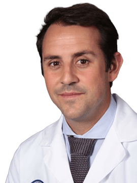
Dr. Gonzalo P. Martin Martin (Cádiz-Spain, 1983) has been working as a General and Digestive System Surgeon in the General Surgery Team of the López-Cano Medical Group in the city of Cádiz since 2023. His academic training as a Bachelor of Medicine was developed at the University of Cadiz and, thanks to international scholarships, he completed his training at the Universitá Cattolica del Sacro Cuore - School of Medicine "A.Gemelli" in Rome and at the Westfälische Wilhelms -Universität Münster - Medizinesche Fakultät in Germany. He specialized in General and Digestive System Surgery under the direction of the renowned surgeon Dr. José Vicente Roig Vila in Valencia and, thanks to obtaining two scholarships as the best curriculum of resident doctors in Valencia and resident surgeons in Spain, he completed his specialist training at the Cleveland Clinic Hospital in the United States, considered one of the best in the world, where he completed his training with the internationally renowned Dr. Steven D. Wexner. In 2015 he received his doctorate with Cum Laude appointment at the historic Faculty of Medicine of the University of Valencia. During the years 2013 to 2019 he was part of the Colorectal Surgery Departments at the Hospital Universitario Son Espases in Palma and at the Hospital San Pablo in Barcelona. In the period from 2020-2022, he was part of the prestigious team of private Surgical Surgery Barcelona, with main activity in the traditional and internationally recognized Teknon Medical Center. During this period, he was also responsible for the Department of Coloproctology and Complex Abdominal Wall at the Sagrada Familia Clinic in Barcelona. Dr. Martin develops his care work with special dedication to colorectal surgery (diseases of the colon, rectum and anus), minimally invasive / endoscopic / laparoscopic surgery, robotic surgery, laser proctology (FACIL technique, etc) and fluorescence-guided surgery; configuring himself as a pioneer in some of these techniques worldwide. In addition to his work in health care, he has served as a professor at renowned universities such as the Universitat Autònoma de Barcelona. As well as coordinator and instructor in training programs in colorectal pathology at national and international level. This intense care and teaching work is accompanied by a passionate research production with more than 100 conference papers, articles in journals of impact, invited lectures such as TEDx conferences and international awards. He has also carried out his scientific work as a junior editor in the journal Techniques in Coloproctology, one of the journals considered to have the highest impact in colorectal surgery and innovation. Similarly, he served on the Board of the Coloproctology Section of the Spanish Association of Surgeons for the years 2019-2022. In 2017 he obtained the recognized title of Fellow of The European Board of Coloproctology which confers him the expert certification in Coloproctology recognized by the European Union. During the same year he was winner of the 2017 innovation grant of the Collegi Oficial de Metges Illes Balears for the development of robotic surgery in the Postdoctoral Robotic Colorectal Surgery Fellow program at the Korea University Anam Hospital in Seoul, considered the capital of robotics, in South Korea.
Selected publications from PubMed
Laser fistula treatment: beyond the controversial aspects: best clinical practice recommendations from an international group of surgeons with extensive experience in the procedure-the FiLaC recommendations.
Ambe PC, Martin-Martin GP, Alam AA, Chaudhri S, Bogdanic B, Ma H, Bolik B, Roman IH, Wu J, Hernandez JDP, Vasas N, Dong Q, Istok P, Schouten R, Kalaskar S, Yao Y, Bruketa T, Koulouteri E, Dobricani V, Zhe C, Giamundo P
Tech Coloproctol. 2025 Jun 9;29(1):131 doi: 10.1007/s10151-025-03164-w.
Fluorescence-based bowel anastomosis perfusion evaluation: results from the IHU-IRCAD-EAES EURO-FIGS registry.
Spota A, Al-Taher M, Felli E, Morales Conde S, Dal Dosso I, Moretto G, Spinoglio G, Baiocchi G, Vilallonga R, Impellizzeri H, Martin-Martin GP, Casali L, Franzini C, Silvestri M, de Manzini N, Castagnola M, Filauro M, Cosola D, Copaescu C, Garbarino GM, Pesce A, Calabrò M, de Nardi P, Anania G, Carus T, Boni L, Patané A, Santi C, Saadi A, Rollo A, Chautems R, Noguera J, Grosek J, D'Ambrosio G, Ferreira CM, Norcic G, Navarra G, Riva P, Quaresima S, Paganini A, Rosso N, De Paolis P, Balla A, Sauvain MO, Gialamas E, Bianchi G, La Greca G, Castoro C, Picchetto A, Franchello A, Tartamella L, Juvan R, Ioannidis O, Kosir JA, Bertani E, Stassen L, Marescaux J, Diana M
Surg Endosc. 2021 Dec;35(12):7142-7153 doi: 10.1007/s00464-020-08234-8.

Aim: Show step by step the gracilis muscle interposition technique in the treatment of complex rectovaginal fistula with the technological incorporation of fluorescence-guided surgery to assess correct flap perfusion. Material and method: A 76-year-old woman with a history of squamous cell carcinoma of the anus was treated with radiotherapy and chemotherapy. Currently disease free. She presents a complex fistula characterized by a diameter greater than 2.5 cm, medium height, and poor quality of the surrounding tissues. A fluorescence-guided tissue transposition technique is performed. With the patient in the lithotomy position, a transperineal approach is performed, with dissection of the rectovaginal space and closure of the affected rectal and vaginal portion. The gracilis muscle is dissected circumferentially through an incision on the inner side of the thigh, preserving its localized vascular supply at the junction of the proximal third with the distal two thirds at the posterior level of the muscle. It is divided above its distal insertion. Finally, it is mobilized to tunnel it subcutaneously to the dissected rectovaginal space, interposing itself between the rectal and vaginal repairs. ICG infusion fluorescence technology is used to assess the correct vascularization of the flap and thus prevent possible causes of necrosis in a fast, safe and simple way. The gracilis muscle is sutured at that level in order to fix it and prevent its retraction. Conclusions: The technical goal is to place healthy, well-vascularized muscle tissue in the rectovaginal space after closure. Meticulous identification and preparation of the vascular pedicle is crucial. Assessment of muscle perfusion after full mobilization and before final placement is critical. And fluorescence-guided surgery is a tool that can help us achieve this goal.
Published
May 2023

Diastasis of the rectus muscles is more common in women due to the puerperium and is associated with midline hernias. Endoscopic surgery makes it possible to restore the linea alba and treat its associated defects by offering a minimally invasive approach as an alternative to the more invasive classic open techniques. Diastasis of the rectus muscles is a secondary separation of these muscles favored by the progressive weakening of the linea alba during pregnancy. Its symptoms range from the feeling of a bulging belly to the presence of midline hernias and pain, among others. The REPA technique consists in restoring the firmness of the abdominal wall through the approximation of the rectus muscles. If there are midline hernias, these are repaired in the same procedure. The endoscopic approach is performed by placing three trocars, usually over the previous caesarean section scar, if there is one.
Published
Oct 2021

Aim: Treatments of pilonidal disease should not be more disabling for the patient than the disease itself. Because of the wide range of surgical options available, the surgeon treating pilonidal disease should master 3–4 approaches that are applicable to a wide range of disease presentations. Simple tract unroofing and curettage are helpful and the most popular option in the setting of minor disease. However, healing time and discomfort are high. Potential advantages of non-excisional therapies are more rapid recovery and less post-procedural pain. The objective of the video is to give a step-by-step description of the laser-based treatment of this frequent proctological pathology. We have named this technique according to our protocol S-FACIL (Sinus-Fistula Ablative Contraction Intracavity Laser). Methods: We present the case of a young man with a diagnosis of standard pilonidal sinus and an indication for treatment in our institution. S-FACIL was applied under spinal anesthesia after a small skin incision of 0.5 cm in every pit with the help of a punch and careful cleaning of the sinus tracts with a curette and peroxide. A radial fiber connected to a diode laser set at the wavelength of 1470 nm was then introduced into the tracts. The laser energy was delivered at 12W with 8-second pulses and 1-second pauses between pulses. The procedure was performed on an outpatient basis with subsequent home cures based on Prontosan®and Aquacel®for 4 weeks. Outcome: The concept of action of this technique is the contraction of the tissue radiated with laser technology in order to eliminate the cavity and avoid a large excisional scar. There are few descriptions of this technique with procedural variations and laser parameters with promising initial results.1 Operative time was 20 minutes with no intra or postoperative complications and the patient achieved complete healing at 6 weeks with minimal deterioration in his daily activity. Conclusion: Potential advantages of modern techniques based on cutting-edge technology like S-FACIL are more rapid recovery and greater comfort.
Published
Sep 2021
Help your network discover Dr. Gonzalo Pablo Martín-Martín's clinical expertise.