Dr. Esteban Emiliani is a urologist and academic recognized worldwide for his discipline in the treatment and prevention of kidney stones.
Dr. Emiliani completed his urology residency at the Puigvert Foundation, attached to the Autonomous University of Barcelona and additionally obtained the title of FEBU - Fellow of the European Board of Urology. This title is a recognition of excellence in the knowledge and practice of urologists at the European level. Subsequently, Dr. Emiliani carried out his subspecialization studies in endourology and medical-surgical treatment of kidney stones in two of the most prestigious institutions worldwide. First, at the Tenon Hospital, attached to the Sorbonne University in Paris, under the tutelage of Professor Olivier Traxer. He then went to the MPUH Hospital in Nadiad (India) where he was under the tutelage of Professor Mahesh Desai. Back in Barcelona, Dr Emiliani obtained his PhD - PhD International, Cum Laude - awarded by the Autonomous University of Barcelona and the Sorbonne University, with a study on the development of laser technologies for the treatment of kidney stones.
Prolific in his academic work, Dr. Emiliani has published more than a hundred articles in indexed urological journals, 12 publications in non-indexed journals and has written four international book chapters. He is a member of the editorial board of the World Journal of Urology, Actas Urológicas Españolas, Turkish Journal of Urology and BMC Urology. In addition, Dr. Emiliani is a member of the most prestigious international institutions shaping the future of urology worldwide.
Selected publications from PubMed
In Vitro Evaluation of Pulsed Thulium:YAG and High-Power Ho:YAG Lasers with Multiple Pulse Modulations.
Bravo-Balado A, Sánchez-Puy A, Izquierdo P, Diana P, Piana A, Stracci D, Arena P, Garcia SG, Kanashiro AK, Millán F, Sánchez-Martín F, Esquinas C, Angerri O, Palou J, Emiliani E
J Endourol. 2026 Aug 12;:8927790261473713 doi: 10.1177/08927790261473713.
Small- and large-calibre ureteroscopes: an updated systematic review and meta-analysis.
Di Bello F, Verri P, Bravo-Balado A, Izquierdo P, Fila L, Kanashiro AK, Gogaj R, Wells E, Traxer O, Angerri O, Emiliani E
BJU Int. 2026 Jul 28 doi: 10.1111/bju.70394.
Real-world DISS practice combined with and without access sheath: insights from the global DISS dataset: a EAU endourology study.
Fuligni D, Gauhar V, Pietropaolo A, Castellani D, Ng CF, Fong KY, Martinez BB, Castillo JG, Zawadzki M, Tsaturyan A, Ragoori D, Gadzhiev N, Malkhasyan V, El Hajj A, Becker B, Ying BLT, Kwok JL, Geavlete P, Geavlete BF, Contreras P, Emiliani E, Rico L, Yuen SKK, Somani B
World J Urol. 2026 Jul 20;44(1) pii: 508. doi: 10.1007/s00345-026-06607-7.
Intrarenal thermal and pressure dynamics during ureteroscopy: an in vitro study.
Bravo-Balado A, Jannello LMI, Zorzi F, Quarà A, Moretto S, Corrales M, Emiliani E, Doizi S, Coste F, Traxer O, Panthier F
BJU Int. 2026 Oct;138(4):722-729 doi: 10.1111/bju.70384.
Comparison of TFL and high-power Ho: YAG lasers with multiple pulse modulations: an in vitro study.
Bravo-Balado A, Sánchez-Puy A, Izquierdo P, Diana P, Piana A, Stracci D, Arena P, Gràcia Garcia S, Kanashiro AK, Millán F, Sánchez-Martín F, Esquinas C, Angerri O, Traxer O, Palou J, Emiliani E
World J Urol. 2026 Jun 18;44(1) doi: 10.1007/s00345-026-06514-x.

Clinical History: 52 yo female patient. The patient was taking topiramate to prevent migraines. No other relevant past medical history besides urolithiasis with previous right ureteroscopy and SWL. After ureteroscopy performed in May 2021, during follow-up the patient continued taking the topiramate (a known cause of urolithiasis) and was diagnosed with a new 1.5 cm stone in the right renal pelvis causing obstruction. It was decided to perform a flexible ureteroscopy with previous stenting. Technique description: After putting the patient under general anesthesia, the patient is placed in a lithotomy position, sterilized and draped. A table is set below the patients left leg, the monitors are set at the patients left side and the C arm in the patients right side. On the table, we place dry gauzes and wet gauzes in a surgical dish. Also in the dish, we open the guidewire, a syringe and scissors. Setting the irrigation bag 40 cm above the patient the surgery begins with the small, 9.5 fr semirigid ureteroscope to perform the initial cystoscopy, and placement of a guide wire in the renal cavities, keeping it as a safety wire. Then a semirigid ureteroscopy is performed to check the ureter for stones and for passive dilation. A second guidewire is placed in the kidney. Then the ureteral access sheath is passed through one of the wires (working wire) keeping the second as a safety wire. The access sheath should be inserted gently without forcing the entry. Then the flexible ureteroscopy with a single use scope (Lithovue) is performed with systematic renal inspection. Once the stone is identified the laser (Lumenis Pulse Moses 2.0) is set, a small 200 micron laser fiber is placed and for dusting, low energy, long pulse and high frequency settings are used. The stone is dusted from the periphery. If breathing movements are intense, episodes of apnea can help stabilize the kidney. After completing the dusting, the basket is inserted, preferring small 1.9 fr baskets to remove a fragment for further analysis. Final stone dusting is performed with the popcorn technique until complete dust is achieved. The access sheath is removed and a ureteral inspection is done at the end of the procedure. A double J stent is placed finally with a bladder catheter. Procedure steps: Cystoscopy. Guidewire placement in the renal cavities and keeping it as a safety wire. Semirigid ureteroscopy and placement of second guidewire. Ureteral access sheath placement. Flexible ureteroscopy with renal inspection. Dusting of the stone until small fragments are achieved. Stone removal with basket. Final stone dusting with popcorn technique. Access sheath removal and ureteral inspection. Double J and bladder catheter placement. Trainings Objectives: Be systematic on the procedure, patient placement and setting. Never force the entry of the instruments. Always plan ahead for possible complications and make choices based on what you could resolve. -Plan the fragmentation technique in order to accomplish a stone free status. Extract a stone fragment for analysis.
With Boston Scientific
Published
Apr 2022
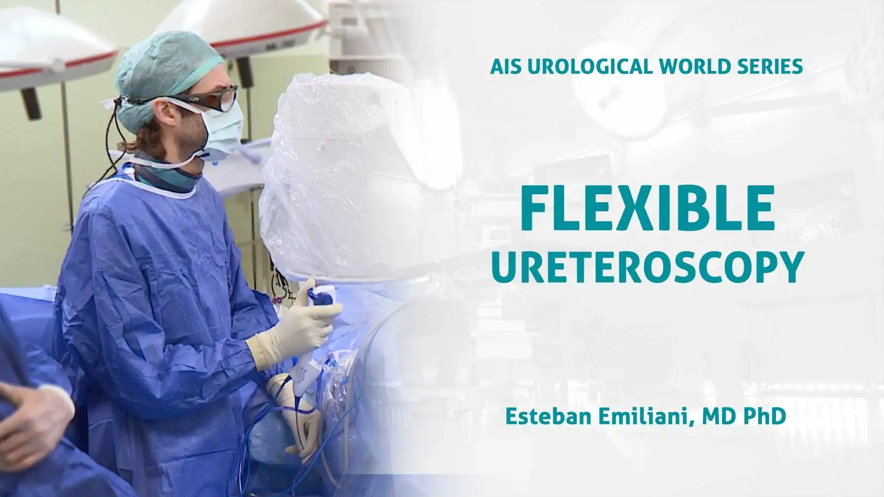
Flexible uteroscopy for renal inspection and stone removal.
With Boston Scientific
Published
Oct 2019
Help your network discover Dr. Esteban Emiliani's clinical expertise.