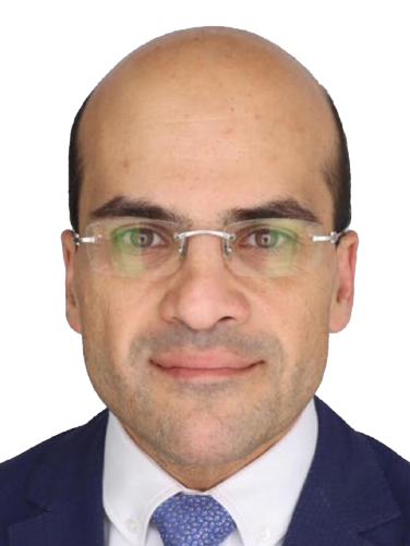
Dr. Abu Dayyeh is Professor of Medicine, Director of Advanced Endoscopy, Director of Metabolic and Bariatric Endoscopy, Vice Chair of Innovation, and Consultant in Gastroenterology and Advanced Therapeutic Endoscopy at the Mayo Clinic in Rochester, MN. He is also the chair of the Bariatric and Metabolic Endoscopy Committee of the International Federation for the Surgery of Obesity and Metabolic Disorders (IFSO) and co-chair of the Bariatric Committee of the American Foregut Society. His research focuses on development of minimally invasive endoscopic solutions for gastrointestinal diseases, obesity, and metabolic disease. Dr. Abu Dayyeh has multiple patents, and he is the founder of a ventures funded start-up company focusing on the endoscopic treatment of diabetes and metabolic disease. Dr. Abu Dayyeh has published more than 200 peer reviewed articles, reviews, and book chapters in the field. His work has been highly cited with more than 3000 citations. He received multiple awards and grants in support of his research, including the Star of Science Award given by His Majesty King Abdulla II during the World Science Forum in the Dead-Sea, Jordan 2017. Dr. Abu Dayyeh has been invited to give many national and international keynote lectures. He serves as a consultant and advisor to multiple leading US and International companies who are developing novel endoscopic therapeutics.
Selected publications from PubMed
A Multicenter Double-blind Randomized Sham-controlled Trial Assessing the EndoBarrier Duodenal-jejunal Bypass Liner for the Treatment of Poorly Controlled Type 2 Diabetes Mellitus With Concomitant Obesity: The ENDO Trial.
Thompson CC, Jirapinyo P, McCarty TR, Brethauer S, Shaheen N, Sullivan SA, Koch T, Abu Dayyeh BK, Wilson EB, Schulman AR, Butsch WS, Gersin KS, Apovian CM, Schauer P
Ann Surg. 2026 Jul 1;284(1):34-42 doi: 10.1097/SLA.0000000000006974.
The Diabetes Remission Index: A Novel Prognostic Calculator Model Predicting Diabetes Remission Before and After Metabolic Procedures.
Ghusn W, Ma P, Vierkant RA, Mundi M, Fehervari M, Ikemiya K, Hage K, Acosta A, Camilleri M, Abu Dayyeh BK, Higa K, Ghanem OM
Ann Surg. 2026 Jul 1;284(1):91-98 doi: 10.1097/SLA.0000000000006656.
A Machine-Learning Assisted Genetic Risk Score Identifies Improved Weight Loss After Endoscopic Sleeve Gastroplasty.
Fredrick T, Maselli D, Vargas E, Wooley C, Gala K, Anazco D, Ciotlos S, O'Connor T, Abu Dayyeh B, McGowan C, Acosta A
Obes Surg. 2026 Jun;36(6):2819-2828 doi: 10.1007/s11695-026-08642-0.
Short-Term Safety and Healthcare Utilization Following Intragastric Balloon Placement in Patients With and Without Diabetes Mellitus: A Propensity-Matched Analysis.
Hamamah S, Samaan JS, Soliman N, Nguyen S, Advani R, Samakar K, Sandhu KK, Park K, Abu Dayyeh BK, Watson RR
Obes Surg. 2026 Apr;36(4):1849-1861 doi: 10.1007/s11695-026-08622-4.
Safety Profile of Primary Intragastric Balloon Placement in Class III Obesity: An MBSAQIP Analysis of 4,555 Patients.
Hamamah S, Samaan JS, Soliman N, Nguyen S, Srinivasan N, Hai F, Samakar K, Sandhu KK, Park K, Abu Dayyeh BK, Watson RR
Obes Surg. 2026 Apr;36(4):1757-1765 doi: 10.1007/s11695-026-08575-8.

1. Welcome and Introduction - Dr. Luis Felipe Cabrera Vargas (Colombia) 2. Residents Worldwide - Testimonials and perspectives 3. The RR: Highlights - Dr. Luis Felipe Cabrera Vargas (Colombia) 4. Industry Perspective- Mr. Rich Merklinger (USA) 5. The Resilient Chain - Dr. Barham Abu Dayyeh (USA), Dr. Julia Coleman (USA), Dr. Zoe Garoufalia (USA), Dr. James Glasbey (UK), Dr. Geeta Lal (USA), Dr. Emanuele Lo Menzo (USA), Dr. Winnie Mathur (India), Dr. Violeta Moizé (Argentina), Dr. Sara Tavares Nogueira (Spain), Dr. Daniel Tomey (USA) 6. Closing remarks - Dr. Luis Felipe Cabrera Vargas (Colombia)
With Johnson & Johnson MedTech
Published
Dec 2022

PROGRAM Welcome and Introduction - Dr. Samuel Szomstein (USA) and Dr. Ashita Jain (India) Leadership challenges and opportunities during residency - Dr. Juan Javier-Desloges (USA) Self-management of leadership and application in the resident program – Dr. Courtney Sommer (USA) How to develop and evaluate a leadership training program for residents – Dr. Barham Abu Dayyeh (USA) Discussion and closing remarks – All faculty moderated by Dr. Ashita Jain (India)
With Johnson & Johnson MedTech
Published
Sep 2022

Flexible gastrointestinal endoscopy started about sixty years ago as a diagnostic discipline. Soon afterward, therapeutic applications were introduced to treat gastrointestinal polyps and bleeding lesions. Sophisticated maneuvers and creative technology permitted endoluminal therapy for tumors of the pancreas and biliary tree. More recently, surgical therapy has been developed for a number of maladies such as obesity, achalasia, and gastroparesis. The use of endoscopic ultrasound has added immensely to the ability of the endoscopist to see and treat numerous extra-luminal conditions. This program, presented by world experts in these new methods, will inform the audience regarding advances and possibilities in these areas.
With Olympus
Published
Jun 2022

CASE 1: Transjejunal laparoscopic-assisted ERCP a technique to deal with choledocholitiasis after a Roux-en-Y reconstruction. The patient is a female of 31 years old that undergone a Roux-en-Y Gastric Bypass two years earlier and presented periodic abdominal pain. MRCP showed stones on the biliary common duct. In the preoperative CT scan no internal hernias were found. TREATMENT: It was decided to perform a laparoscopic-assisted ERCP. The first thing to do was to identify the gastro-jejunal anastomosis and the jejunal-jejunal anastomosis. An internal hernia in the Petersen space was found but immediately reduced and closed. Then the cholecystectomy was performed. Once the limb near the Treitz ligament was identified and pulled through a mini-laparotomy, a colonoscope was introduced and a plastic stent was placed in the papilla, which was the reference for the sphincterotomy. After doing that, a 5mm stone came out. Then the bile duct was checked with a fogarty balloon catheter and a cholangiogram. Then enterotomy was closed and it was checked for any biliary lesions. OUTCOME: After the surgery the patient started liquid diet the first day and was dismissed after 4 days. Questions were about the use of colonoscope rather than duodenoscope, absorbable suture, mini-laparotomy and transgastric and totally endoscopic approach for similar cases. CASE 2: Endoscopic tunneled stricturotomy with full-thickness dissection in the management of a sleeve gastrectomy stenosis. The patient was a 28 years old women with a history of a sleeve gastrectomy in 06/2018. (BMI: 35.3) who was presenting dysphagia to solid food. During the progression to solid food she developed food regurgitation and vomiting and that is why upper-GI series were performed (stenosis was identified). To treat this stenosis she underwent 3 endoscopic pneumatic balloon dilatations. Those were not effective and she lost 30kg in 5 months so it was decided to perform an endoscopic tunneled stricturotomy with full-thickness dissection. TREATMENT: To start the procedure, once the stenosis was identified a submucosal injections is performed 3-5 cm before the stenotic area. Then, an incision was performed to tunnel submucosally. In this part of the surgery is important to stay on the submucosal layer and not go full-thickness. Then a myotomy was performed and in the area marked with both yellow arrows (stapled line) we dissect out performing the full-thickness stricturotomy. After that, we close using clips. We can see a comparison before and after performing the procedure showing a much larger lumen. OUTCOME: The patient had no post-procedure symptoms and during follow-up the patient tolerated diet and no recurrence of the symptoms appeared. At 2 months, we can see the differences in the upper-GI series and in the endoscopy. As a conclusion endoscopic tunneled stricturotomy with full-thickness dissection appears to be safe and effective in the management of stenosis after sleeve gastrectomy. This procedure can be used after conventional techniques such as pneumatic balloon dilatation or stents fail, or may be considered as an initial therapy for sleeve stenosis. Discussion was based on patient selection, indications, myotomy technique and leak rates. CASE 3: From Nissen Fundoplication to RYGB to treat both GERD and morbid obesity Patient was a 31 year-old woman that had an open Nissen fundoplication on 2010 and a surgical revision with a redo of it on 2012. She complained with dysphagia and morbid obesity so an extensive preoperative workout was done. In the Barium swallow, an esophageal dilatation with a cardial stenosis was presented, and also, an additive image on the left side of the esophagus suggestive of an ulceration. The Upper-GI endoscopy revealed an esophagitis grade C and the manometry showed incomplete waves at lower 3rd and inappropriate release of LES. In the pH-metry acid reflux and positive DeMeester score were evidenced. TREATMENT: The procedure starts dissecting the perigastric tissue, that is challenging as it presented high fibrotic component. Then the dissection of the gastric valve and the hiatal region from the right to the left was completed. Once that was performed an small gastric pouch of 20cc was created to reduce the acid production. Then as in a normal RYGB the biliopancreatic is done at 50cm and the alimentary at 150cm. Jejuno-jejunal and gastro-jejunal were handsewn. OUTCOME: No events were presented in the postoperative, patient at two years follow-up has a BMI of 27, No GERD and no IPP. As a conclusion RYGB is a great option to treat GERD in obese patients. We cannot forget dysphagia as a complication of Nissen fundoplication and conversion from Nissen to RYGD allows us to treat dysphagia, GERD and obesity. Different topics were discussed after the presentation of this case as: short gastric pouch, differences with regular bypass, situations where you cannot undo the Nissen fundoplication, mesh indications above others. CASE 4: From RYGB to SADS Patient was a 49 year-old woman with an initial BMI of 57 and it was decided to convert to SADS for weight regain. TREATMENT: The first thing to do was remove all adhesions from previous surgeries and then dissect the Hiss angle. Once this has been done, he identifies the Roux limb and dissects it 25cm after the gastric pouch. The next thing to do was to connect the greater curve with the Hiss angle. He disconnects the previous anastomosis and performs a gastro-gastrostomy. It is important to do it near the lesser curve as after that a Sleeve gastrectomy will be performed using a Bougie of 42 frames as a reference. The transection of the duodenum is performed conserving as much vascularization as it is possible in order to maintain sleeve well irrigated. After that duodenum-ileal anastomosis handsewn was performed. Anastomosis were checked with methylene blue. OUTCOME: The outcome was good and the patient was discharged 2 days after the procedure with no complications. Questions were about rate of complications, length of limbs, roux limb, conversion after RYGB and weight loss after conversion. CASE 5: PETERSEN’S HERNIA AFTER OAGB Patient was a 48 years-old woman with a BMI of 52, she was referring bile reflux with no response to medical treatment. So it was decided to convert mini-gastric bypass to RYGB. TREATMENT: As the surgery started, an internal hernia was identified on the Petersen’s defect, in this case the hernia was protruded from medial to lateral something that is rare as normally it is the other way around. To start the procedure we need to identify the proximal and distal limb of gastrojejunal anastomosis. On the proximal limb of this anastomosis an enterotomy was performed and 75cm of the alimentary limb were measured to perform the jejunojejunal anastomosis. After that the jejunojejunal and Petersen’s defect were closed. After doing that, the transection of biliopancreatic limb from alimentary limb was performed. OUTCOME: The outcome was good and the patient was discharged 1 day after the procedure with no complications. A short term follow-up at 3 months was symptoms free. In this last case, discussion was based on rate bile reflux, biliopancreatic limb length, indications for conversion, causes for bile reflux and hernia defect closures.
Published
Feb 2020

Mild to moderate obesity is an entity with associated complex morbidity. However, these patients may not be candidates for surgical treatment. In these cases, endoscopic procedures such as intragastric balloons, gastric remodeling/plication, gastric aspiration therapy, bypass devices, bile diversion, or balloon occluded gastrojejunostomy may be considered. Balloons may result in greater weight loss versus placebo when supported by an adequate follow-up and dietary recommendations. The national registry of use of balloons in the United States is presented, as well as the international data. Sequential balloon placement results are also discussed. The gastric remodeling or plication involves an endoscopic sleeve gastroplasty, created with a suturing endoscopic instrument in an outpatient setting. Results show that it is a quick and safe procedure based on the prospective studies. In cases of weight regain, it can be repeated endoscopically. Gastric aspiration therapy is applied following the same technique as PEG placement, and it can stay for up to 5 years. It achieves weight loss and also helps patients change their eating behavior. Another newer technique involves creating an endoscopic anastomosis, building a partial biliopancreatic diversion, or a gastrojejunostomy created endoscopically. [Image]
Published
Apr 2018
Help your network discover Dr. Barham Abu Dayyeh's clinical expertise.